
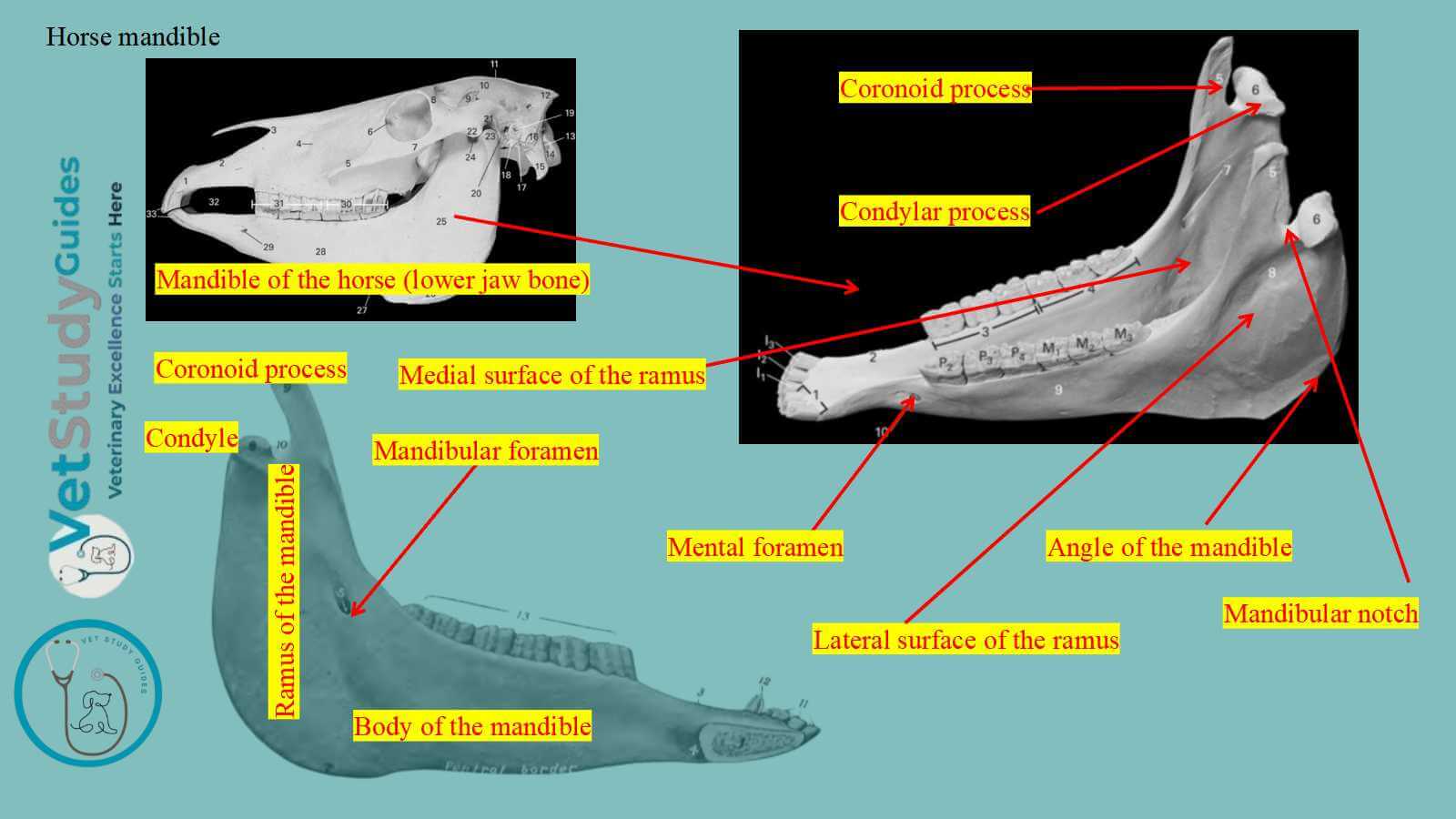
The horse mandible forms the lower jaw. However, the mandible is the largest bone of the horse’s face.
In this guide, I will describe the horse’s mandible in detail with diagrams.
How is the horse’s mandible formed?
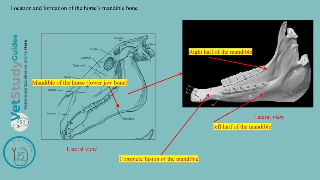
The two halves of the horse’s mandible unite at birth and during the second or third month. However, it is usually described as a single bone in the horse skull.
Again, it carries the lower teeth and articulates its condyles with the squamous temporal on either side. Here, the figure shows how the horse’s mandible forms.
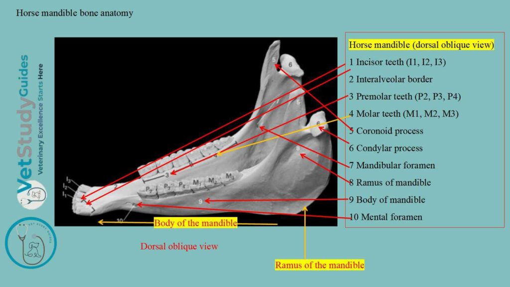
Horse mandible bone anatomy
For descriptive purposes, the horse’s mandible consists of a body and two rami. Here, the body of the mandible is the horizontal part, whereas the rami are the vertical parts of the bone.
To describe the anatomy of the horse’s mandible, you need to describe the osteological features of both parts. Here, I will describe the features of the body and rami of the equine mandible, with diagrams shown separately.
Body of the equine’s mandible
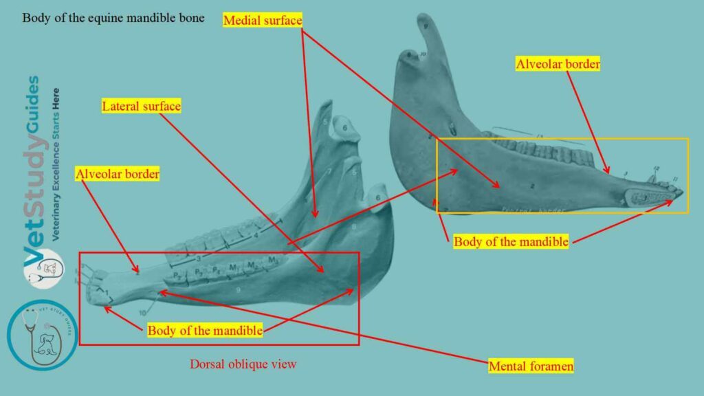
The body of the mandible, or corpus mandibulae, is the thick anterior part that bears the incisor teeth. It presents two surfaces and a border.
Here, the body has two surfaces: the lingual surface and the mental surface.
Again, the border is the curved alveolar border.
Surfaces of the mandibular body
Lingual surface: The lingual surface, or facies lingualis, is smooth and slightly concave. However, during life it is covered by mucous membrane, and the tip of the tongue overlies it.
Mental surface: Again, the mental surface, or facies mentalis, is convex and related to the lower lip. It is marked by a median furrow which indicates the position of the primitive symphysis mandibulae.
Border of the body of the equine’s mandible
The curved alveolar border, or limbus alveolaris, presents six alveoli for the incisor teeth. However, a little further back two alveoli for the canine teeth in the male. In the mare, the alveoli for the canine teeth are usually absent or small.
Rami of the horse’s mandible
The rami of the horse’s mandible extend backward from the body and diverge to enclose the mandibular space. Each ramus is bent to consist of a horizontal part and a vertical part.
- Horizontal part: It bears the lower cheek teeth,
- Vertical part: It is expanded and furnishes attachment to powerful muscles.
However, the term angle is applied to the most prominent part of the curve between these two parts.
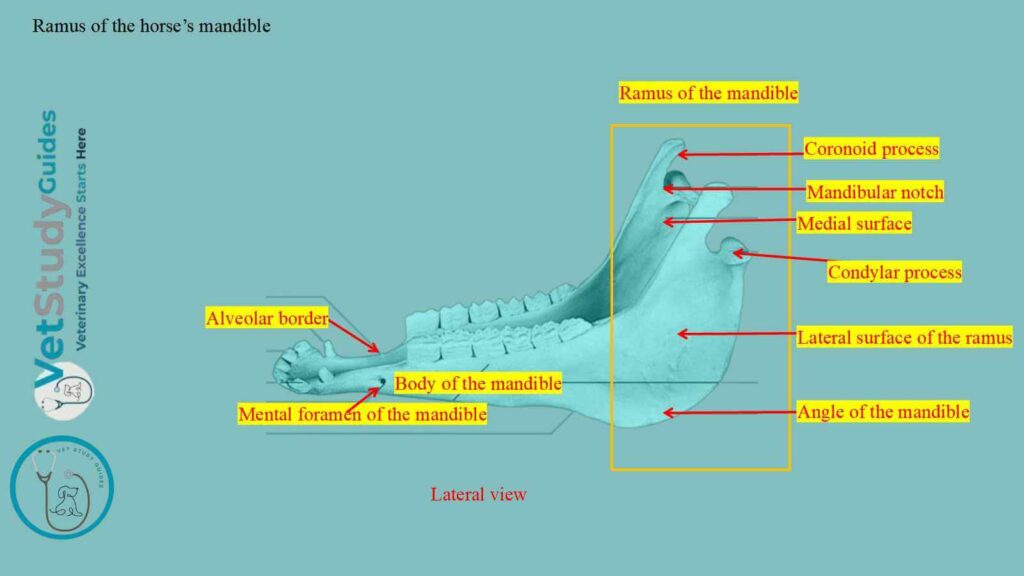
The ramus of the horse mandible presents two surfaces, two borders, and two extremities.
- Surfaces are: lateral and medial.
- Borders are: dorsal and ventral.
- Extremities are: anterior and articular.
Surfaces of the rami of the equine mandible
Lateral surface: The lateral surface is smooth and slightly convex from edge to edge on the horizontal part. At the junction of this surface with the body, it presents the mental foramen.
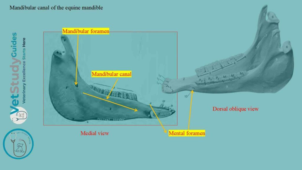
However, the mental foramen is the external opening of the mandibular canal. On the vertical part, it is somewhat concave and has several rough lines for attachment of the masseter muscle.
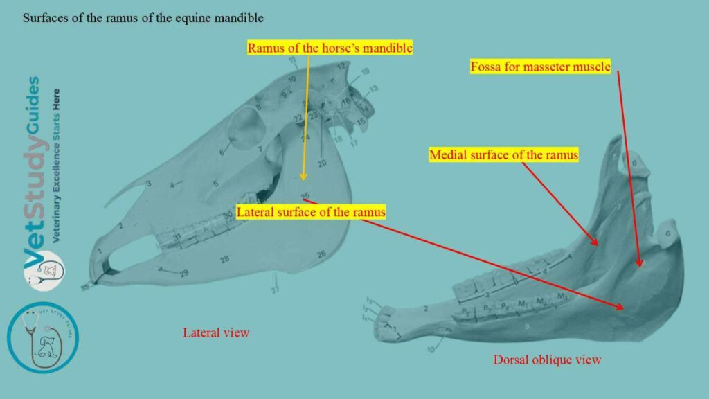
Medial surface: The medial surface of the horizontal part is smooth and presents a shallow longitudinal depression in its middle. Above this, there is often a faint mylohyoid line for attachment of the mylohyoideus muscle.
At the lower part of the junction with the body there is a small fossa. However, this fossa is for the attachment of the genio-hyoid and genioglossus muscles.
Again, on the vertical part, the medial surface is concave. However, its lower and posterior part is marked by rough lines for the attachment of the horse’s medial pterygoid muscle.
Mandibular canal: In front of the middle (pterygoid muscle) is the mandibular foramen, the posterior orifice of the equine’s mandibular canal. However, the canal curves downward and passes forward below the horse’s cheek teeth, opening externally at the mental foramen.
However, it continues into the body of the bone as a small canal. This canal carries the vessels and nerves to the incisor teeth.
Borders of the rami of the horse’s mandible
Dorsal border: The dorsal or alveolar border forms the anterior part of the interalveolar space. Here, this dorsal border is thin.
Behind this it is thick and is excavated by six alveoli for the lower cheek teeth. Again, behind the last alveolus it curves sharply upward and is narrow and rough.
In the young foal, a small alveolus for the vestige of the first premolar is commonly present close to the first large one. This is also known as the wolf tooth in the young foal.
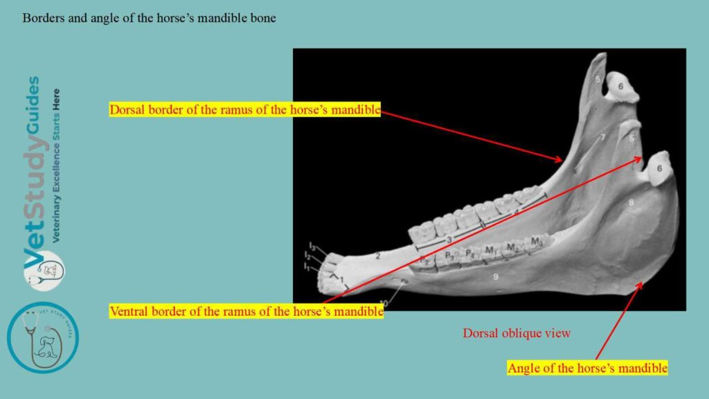
Ventral border: The ventral border of the horizontal part is nearly straight. However, it is thick and rounded in the young horse. Again, it becomes narrower and sharper in old subjects.
At its posterior part, a smooth impression forms where the facial vessels and parotid duct turn around the bone.
Angle of the mandible: Behind the smooth impression, the border curves sharply upward, forming the angle of the mandible. This part is thick and has two roughened lips, separated by a considerable intermediate space. However, near the condyle, it becomes narrower.
Extremities of the rami of the equine mandible
Anterior extremity: The anterior extremity of the rami of the horse’s mandible joins the body.
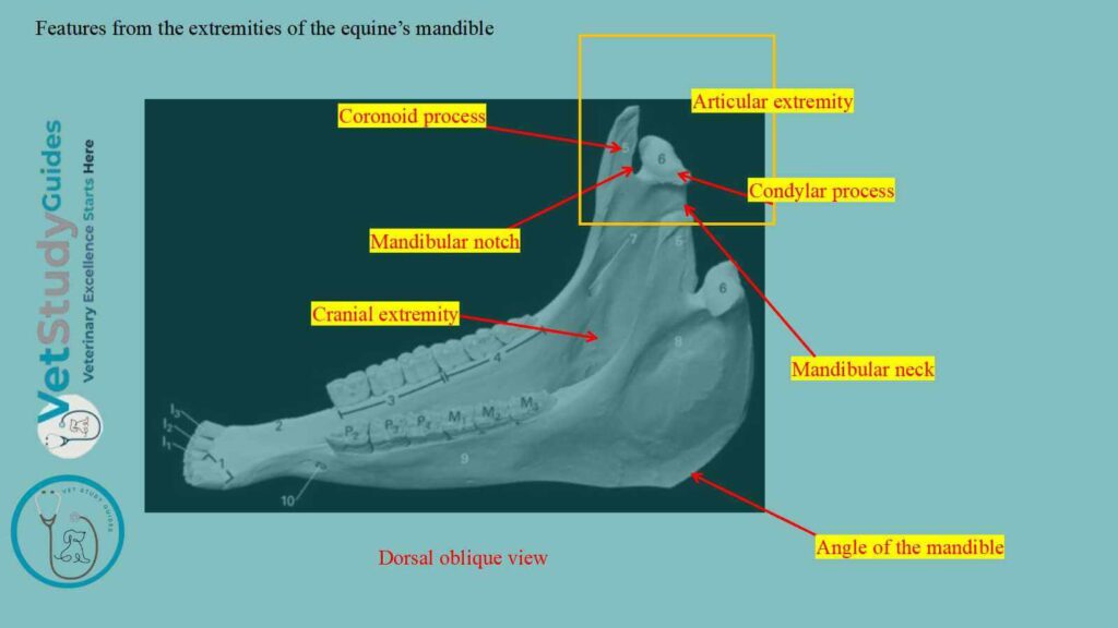
Articular extremity: The articular extremity comprises the coronoid process in front and the condyle behind. However, the mandibular notch separates these two structures. Within this notch, the nerve passes to the masseter muscle.
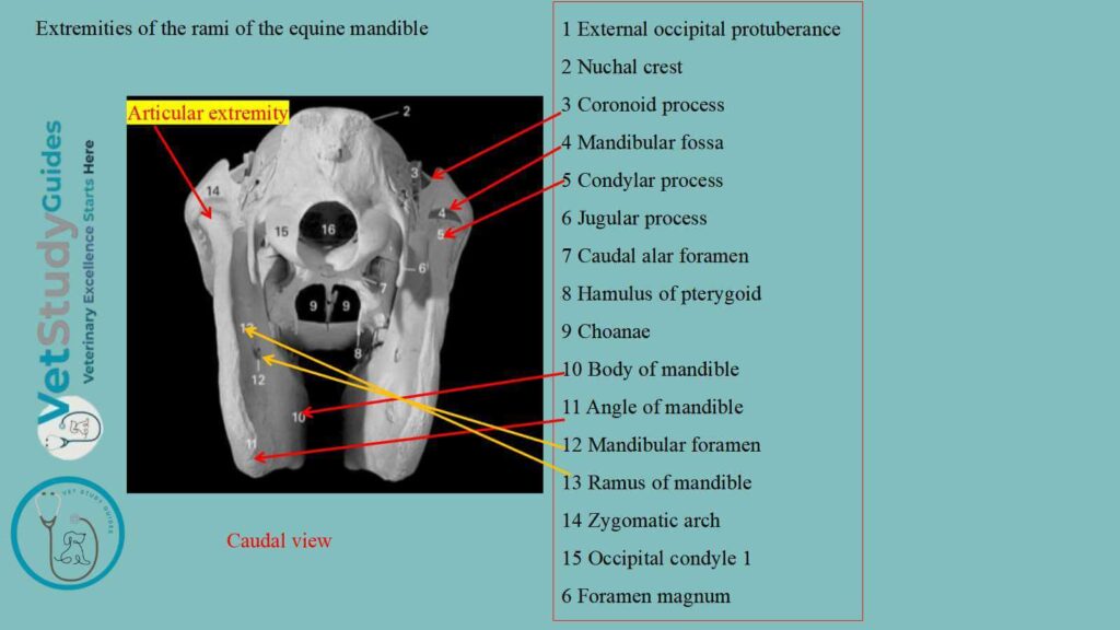
Coronoid process: The coronoid process is thin transversely and curved slightly medially and backward. It projects upward into the temporal fossa and provides an insertion for the temporalis muscle.
Condylar process (condyle): The condyle of the mandible lies much lower than the end of the coronoid process. It is elongated transversely and articulates with the squamous temporal through an articular disc.
Neck of the mandible: The part below the condyle is usually termed the neck of the mandible. On its antero-medial part is a depression, the fovea pterygoidea. Within it, the lateral pterygoid muscle is attached.
The middle of the vertical part of the ramus consists to a large extent of a single plate of compact substance. This structure may be so thin in places that it is translucent.
How does the horse’s mandible change with age?
These changes are largely associated with the growth, and later the reduction, of the teeth. In the young horse, the teeth are long and largely embedded in the mandible.
Here, the body is thick and strongly curved, and the horizontal part of the ramus is also thick. Later, as the teeth are extruded from the bone, the body becomes flattened and narrower.
Again, the horizontal part of the ramus is thinner, especially in its lower part.
In the old subject, the angle and the vascular impression in front of it are developed. However, the lines for the attachment of the tendinous layers of the masseter and pterygoid muscles are more pronounced.
FAQ’s on the horse’s mandible
The equine mandible develops from two chief centers in the connective tissue overlying the paired Meckel’s cartilages. At birth, the equine’s mandible consists of two symmetrical halves which meet at the median symphysis mandibulae. Fusion usually occurs in the second or third month of age.
The horse’s mandible bone is also commonly called the inferior maxilla. Here, the body bears the teeth and is divided into a pars incisiva and a pars molaris. Again, the ramus is the rest of the bone.
Conclusion
So, the two halves of the mandible form the single mandible in the horse. However, it consists of the body and rami, with various surfaces, borders, and extremities.
The body of the equine/horse mandible contains the alveolar sockets, mental foramen, and mandibular canal. Again, the rami of the mandible include the coronoid and condylar processes, mandibular notch, and the angle.
References
- Sisson, S., Anatomy of the domestic animals. W. B. Saunders Company, USA.
- Dyce and Wensing, Textbook of Veterinary Anatomy, 4th edition, Saunders, USA.
- Carmalt and Pimentel (2022). The Equine Temporomandibular Joint. Frontiers in Veterinary Science, 9, 876041.
- Beth Vanhorn and Robert W. Clark, Veterinary Assisting: Fundamentals & Applications, ISBN-13: 978-1-4354-5387-6, Maxwell Drive, Clifton Park, NY 12065-2919 USA.
- Anna Dee Fails and Christianne Magee, Anatomy and physiology of the Farm Animals, 111 River Street, Hoboken, NJ 07030, USA.
- Fewson, S., et al. (2026). The Peridental Structures of Equine Cheek Teeth. Animals, 16(14), 222
- Hilary M. Clayton, Peter F. Flood, Diana S. Rosenstein, and David Mandeville, Clinical Anatomy of the Horse, First edition 2005, ISBN 07234 3302 X.
- Pasquini and Spurgeon, Anatomy of domestic animals, systemic and regional approaches.
- Victoria Aspinall B, and Melanie Cappello, Introduction to Veterinary Anatomy and Physiology Textbook, ISBN 978-0-7020-5735-9, Elsevier.
- Sub, F., et al. (2024). Gross Anatomy of the Equine Masseter Muscle. Anatomia, Histologia, Embryologia, 53(6), e70000.
Reviewed and Edited by