The horse patella is a roughly triangular sesamoid bone. In this guide, you will learn the osteological features of the horse’s patella with a diagram.
I will help you to identify all the osteological features of the horse’s patella from the real sample.
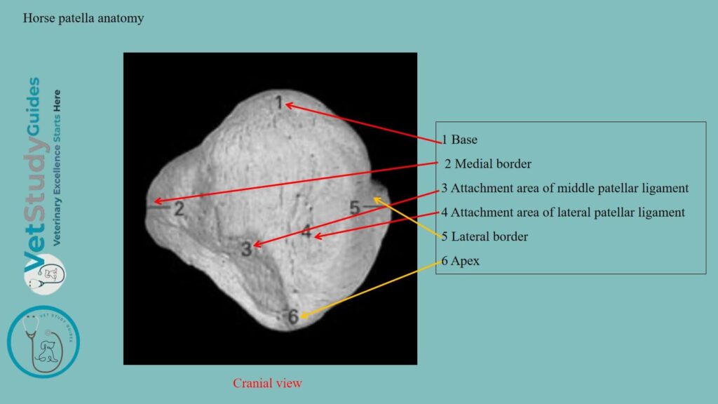
Horse patella anatomy
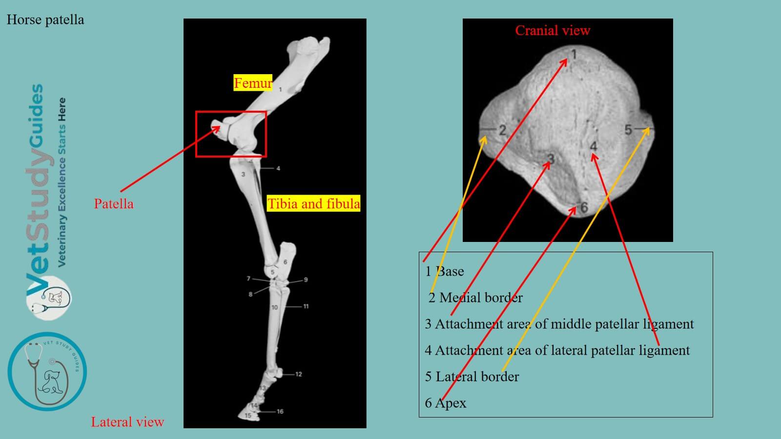
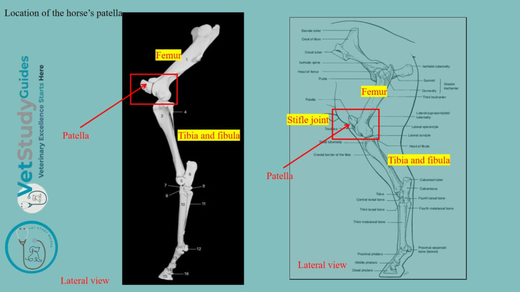
Location of the patella: The patella is a large sesamoid bone which articulates with the trochlea of the femur. Here, the figure shows the location and features of the horse’s patella bone.
Unique features of the horse’s patella bone
Followings are the unique osteological features of the horse’s patella bone –
- This bone is not so triangular in shape,
- The base is narrow, and the apex is blunt,
- Here, the medial and lateral borders of the horse’s patella bone meet at the base at a wide angle,
- The medial and lateral angles are not prominent in the horse’s patella bone,
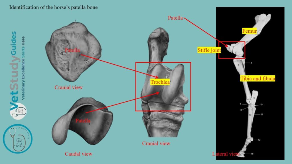
Identification of the horse’s patella bone from its hind limb
Here, the figure shows the patella bone in the front part of the horse’s stifle joint. Thus, to identify this patella bone from the horse skeleton, you might first find the hindlimb bones. In the hindlimb of a horse, you will find the hip bone, femur, tibia and fibula, tarsals, metatarsals, and phalanges (digits).
However, the joints from the horse’s hindlimb are the hip joint, stifle joint, tarsal joint, fetlock, pastern, and coffin joint.
Now, you need to observe the distal end of the horse’s femur and the proximal end of the tibia and fibula bones. Together, these bone along with the patella on the front aspect forms the stifle joint.
Now, you need to identify the different osteological features of the horse’s patella. For this, the free area (in front) of the patella is the free or anterior surface.
Again, the attached area with the trochlea of the femur is the attached or articular surface.
Now, the area or border that faces dorsally is the base of the horse’s patella. However, the blunt point that faces ventrally is the apex of the horse’s patella.
Table 1 presents the features that you need to identify from the horse’s patella bone –
| Surfaces/borders/others | Features |
| Surfaces | Anterior and articular surfaces |
| Borders | Lateral and medial borders |
| Base | Dorsally wide but narrow than the ox |
| Apex | Ventrally directed blunt area |
Description of the horse patella bone
Here, the horse’s patella presents for description 2 surfaces (anterior, posterior), 2 borders (lateral, medial), a base, and an apex.
- Surfaces: anterior and posterior (articular)
- Borders: lateral and medial border,
- Base: It faces upward and backward, and
- Apex: It faces ventrad and forms the blunt point
Anterior surface of the horse patella
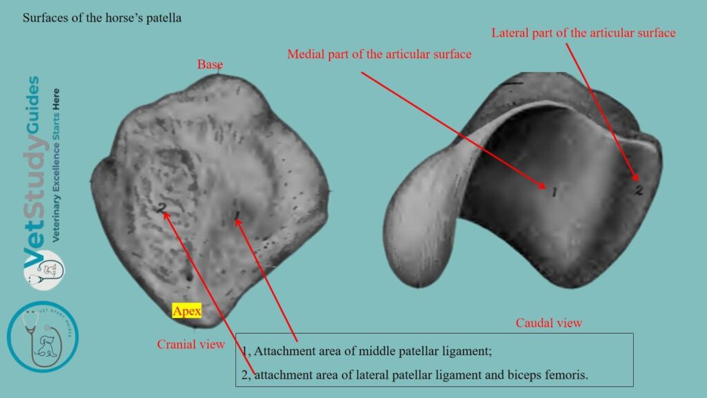
The anterior is also known as the free surface/facies libera. It is a quadrilateral, convex, and rough for muscular and ligamentous attachment.
Articular surface of the patella bone
The articular surface or facies articularis is also quadrilateral. But this surface is much less extensive.
It presents a vertical, rounded ridge, which corresponds to the groove on the trochlea of the horse’s femur. However, it separates two concave areas.
Of the latter, the medial one is much the larger. It is not very well adapted to the corresponding ridge of the trochlea of the femur in the fresh state.
However, it is completed and rendered more congruent by the curved accessory fibro-cartilage.
Borders of the horse’s patella bone
The borders are medial and lateral, and converge to the apex below. Together, these borders form an angle at the base of the patella bone.
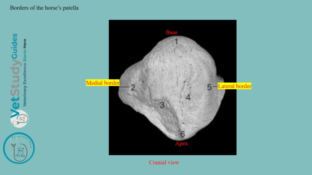
Medial border: The medial border is concave.
Lateral border: The lateral border is rounded in the horse’s patella. Its angle is less prominent than the medial.
The medial angle and the adjacent part of the posterior margin of the base give attachment to the fibro-cartilage of the patella.
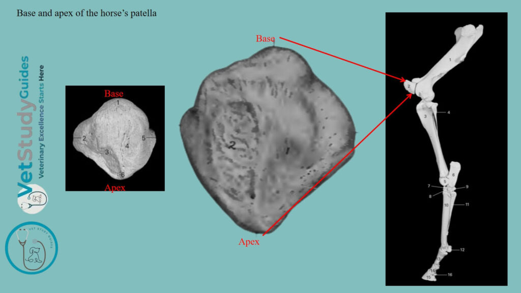
Base and apex of the horse’s patella
The base or basis patellae faces upward and backward. It is convex transversely, concave from before backward.
However, the apex or apex patellae forms a blunt point directed distally.
FAQ’s on the horse patella bone anatomy
Well, the horse’s patella develops as a sesamoid bone from a single center in a cartilaginous structure. It is deposited in the tendon of the quadriceps femoris muscle.
The pig patella is different from the horse’s, as it has three surfaces. However, the pig’s patella is smaller than that of the horse’s patella.
Conclusion
So, the horse patella is a triangular sesamoid bone having 2 surfaces, 2 borders, a base, and an apex. It is located in front of the trochlea of the horse’s femur bone.
The base is directed upward and narrow, whereas the apex of the horse’s patella faces ventrally and blunt.
References
- Sisson, S., Anatomy of the domestic animals. W B Saunders Company, USA.
- Dyce and Wensing, Textbook of Veterinary Anatomy, 4th edition, Saunders, USA.
- Tnibar, A. (2003), Treatment of upward fixation of the patella in the horse: an update. Equine Veterinary Education, 15: 236-242.
- Beth Vanhorn and Robert W. Clark, Veterinary Assisting: Fundamentals & Applications, ISBN-13: 978-1-4354-5387-6, Maxwell Drive, Clifton Park, NY 12065-2919 USA.
- Fjordbakk, et al., The equine patellar ligaments. BMC Vet Res 19, 20 (2023).
- Anna Dee Fails and Christianne Magee, Anatomy and physiology of the Farm Animals, 111 River Street, Hoboken, NJ 07030, USA.
- Hilary M. Clayton, Peter F. Flood, Diana S. Rosenstein, and David Mandeville, Clinical Anatomy of the Horse, First edition 2005, ISBN 07234 3302 X.
- DYSON, S.J. (2002), Normal ultrasonographic anatomy and injury of the patellar ligaments in the horse. Equine Veterinary Journal, 34: 258-264. https://doi.org/10.2746/042516402776185976
- Pasquini and Spurgeon, Anatomy of domestic animals, systemic and regional approaches.
- Ludwig, et al., (2023). Bilateral patellar aplasia in a foal. Vet med and sci, 9(3), 1143–1148.
- Dyson SJ. Normal ultrasonographic anatomy and injury of the patellar ligaments in the horse. Equine Vet J. 2002 May;34(3):258-64. https://doi.org/10.2746/042516402776185976
- Busschers, E. (2009). Patellar luxation in horses: Treatment and prognosis. Equine Veterinary Education, 21(9), 464–466.
- Victoria Aspinall B, and Melanie Cappello, Introduction to Veterinary Anatomy and Physiology Textbook, ISBN 978-0-7020-5735-9, Elsevier.
Reviewed and Edited by