
The horse hip bones consist of both the right and left ilium, ischium, and pubis. Here, I will describe the osteological features of the horse’s hip bones with diagrams.
I will describe the ilium, ischium, and pubis bones from the horse hind limb separately.
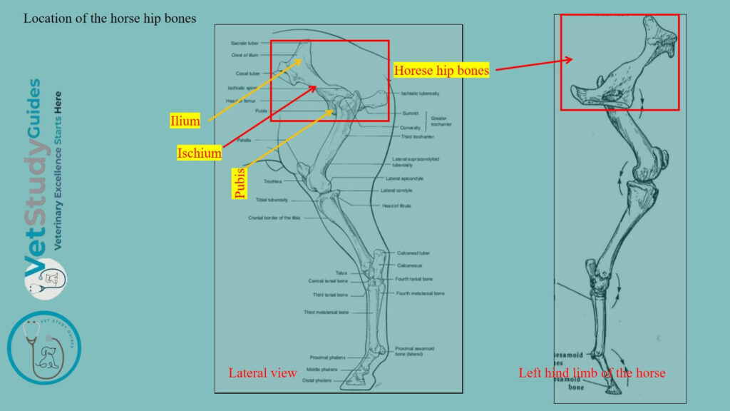
Horse hip bones
The figure shows the location and different osteological features of the horse’s hip bones.
What is the pelvic girdle in a horse?
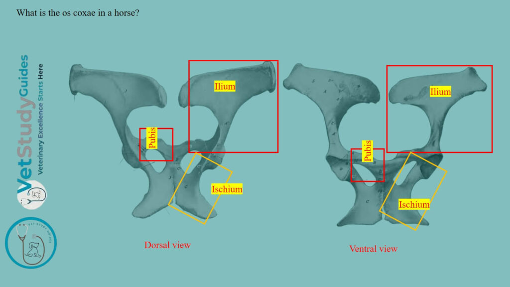
The horse’s pelvic girdle consists of the os coxae. It unites ventrally with the opposite bone at the symphysis pelvis. However, they articulate with the sacrum bone dorsally.
What is the os coxae in a horse?
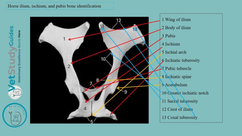
The Os coxae or hip bone is the largest of the flat bones of the horse skeleton. It consists primarily of three parts: the ilium, ischium, and pubis.
They meet to form the horse’s acetabulum. It is a large cotyloid cavity for articulation with the head of the horse femur.
These parts are fused at about one year of age, but it is convenient to describe them separately. Let’s describe these os coxae bones from the horse’s hind limb.
Horse ilium bone anatomy
The ilium, or os ilium, is the largest of the three parts. It is irregularly triangular and presents two surfaces, three borders, and three angles.
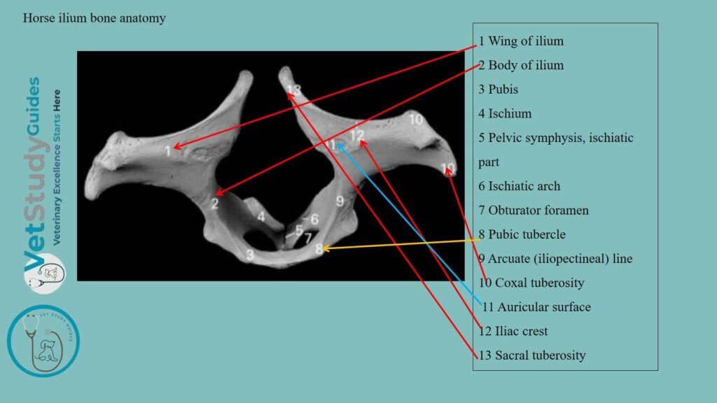
Wing of the horse’s ilium
The wide part of the bone is the wing or ala ossis ilium.
Gluteal surface: Its gluteal surface faces dorso-laterally and backward.
It is wide and concave in front, narrower and convex behind. The curved gluteal line crosses the wide part.
This line extends from the middle of the medial border toward the tuber coxae. However, this surface gives attachment to the middle and deep gluteal muscles.
Pelvic surface: The pelvic surface faces in the opposite direction. It is convex and consists of two distinct parts.
Medial part: The medial triangular part is roughened for ligamentous attachment. It bears an irregular facet, the auricular surface for articulation with the sacrum.
Lateral part: The lateral quadrilateral part is, in general, smooth. It is crossed by the ilio-pectineal line, which begins below the auricular surface.
However, the line is continued on the shaft of the bone to join the anterior border of the pubis. The line is interrupted by furrows for the iliaco-femoral vessels.
There is the psoas tubercle, which gives attachment to the psoas minor muscle. The iliacus muscle is attached to the surface lateral to the ilio-pectineal line.
Borders of the horse ilium bone
Anterior border: The anterior border or crest is concave, thick, and rough. Medial border: The medial border is deeply concave. Its middle part forms the greater sciatic notch, and it is continuous behind with the ischiatic spine.
Lateral border: The lateral border is concave and, for the most part, rough. Grooves cross its anterior part for the ilio-lumbar vessels, which are continued on the pelvic surface.
The nutrient foramen is usually situated on or near the posterior part of this border.
Angles of the horse ilium bone
Medial angle: The medial angle is termed the tuber sacrale. It curves upward and a little backward, opposite to the first sacral spine.
However, this angle (tuber sacrale) forms here the highest point of the skeleton. It is somewhat thickened and rough.
Lateral angle: The lateral angle is the tuber coxae, which forms the basis of the point of the hip. It is a large quadrangular mass, narrow in its middle.
However, it is enlarged at either end, where it bears a pair of tuberosities. It is roughened for muscular attachment.
Acetabular angle: The acetabular angle meets the other two bones at the acetabulum. Its prominent dorsal border forms part of the ischiatic spine, which is roughened laterally and smooth medially.
Shaft of the horse ilium
Two depressions above and in front of the acetabulum give attachment to the tendons of origin of the rectus femoris muscle. The acetabular angle is connected with the wing or wide part of the bone by a constricted part, termed the shaft. The latter is of a three-sided, prismatic form.
Lateral surface: Its lateral surface is convex and rough, and gives attachment to the deep gluteus muscle.
Pelvic surface: Its pelvic surface is smooth and is grooved for the obturator vessels and nerve.
Ventral surface: Its ventral surface is crossed by vascular grooves. Below this groove, there is a rough area, which is bounded medially by the psoas tubercle.
Horse ischium bone anatomy
The ischium or os ischii forms the posterior part of the ventral wall or floor of the bony pelvis. It slopes a little downward and inward. But it is practically horizontal in the longitudinal direction.
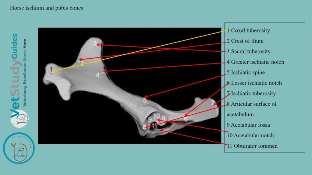
Body of the horse’s ischium
The body of the horse ischium/corpus ossis ischii is irregularly quadrilateral. It may be described as having two surfaces, four borders, and four angles.
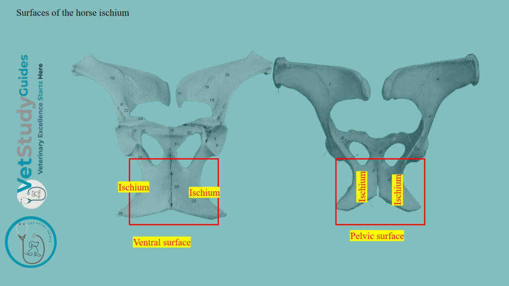
Surfaces of the body of the horse ischium
- Pelvic surface: The pelvic surface is smooth and slightly concave from side to side.
- Ventral surface: The ventral surface is nearly flat and is, in great part, roughened for the attachment of the adductor muscles.
Borders of the horse ischium bone
- Anterior border: The anterior border forms the posterior margin of the obturator foramen.
- Posterior border: The posterior border is thick and rough. It slopes medially and forward to meet the border of the other side, forming with it the ischial arch.
- Medial border: The medial border meets the opposite bone at the symphysis ischii.
- Lateral border: The lateral border is thick and rounded, but concave in its length. It forms the lesser sciatic notch, the lower boundary of the lesser sciatic foramen.
Angles of the horse’s ischium bone
Antero-medial angle: The antero-medial angle or symphyseal branch meets the pubis, with which it forms the medial boundary of the obturator foramen.
Antero-lateral angle: The antero-lateral angle or acetabular branch joins the other two bones at the acetabulum. Dorsally, it bears part of the ischiatic spine, and medially, it is grooved for the obturator vessels.
Postero-medial angle: The postero-medial angle joins its fellow at the symphysis.
Posterolateral angle: The posterolateral angle is a thick, three-sided mass and is termed the tuber ischii. Its lower border is the ventral ischiatic spine, to which the biceps femoris and semitendinosus muscles are attached.
Horse pubis bone anatomy
The pubis/os pubis is the smallest of the three parts of the os coxae. It forms the anterior part of the pelvic floor, and may be described as having two surfaces, three borders, and three angles.
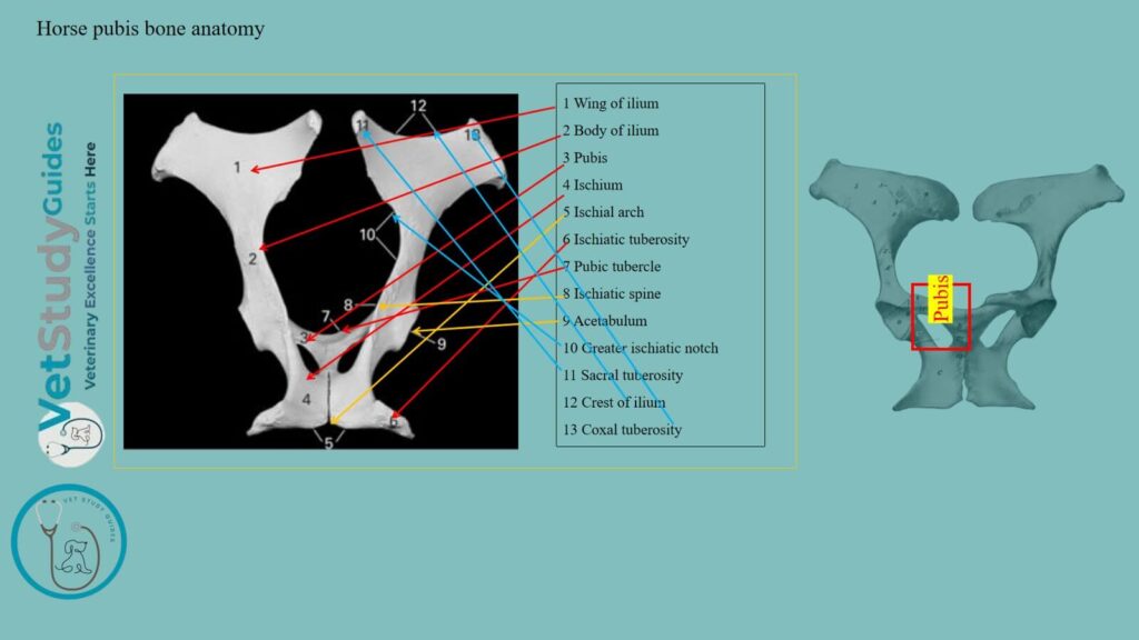
Surfaces of the horse pubis bone
- Pelvic surface: The pelvic surface is convex in the young subject and the stallion. However, it is concave and smooth in the mare and usually in the gelding also.
- Ventral surface: The ventral surface is convex, and in great part rough for muscular attachment. Near the anterior border, it is crossed by the pubic groove.
A large vein occupies the medial part of this surface, and the accessory ligament occupies the lateral part.
Borders of the horse pubis bone
Anterior border: The anterior border is thin in its medial part except in the young subject and the stallion. It forms the pecten ossis pubis in the horse hip bones.
Laterally, it bears the rough ilio-pectineal eminence, beyond which it is continuous with the ilio-pectineal line. Near the symphysis is a variable prominence, the tuberculum pubicum.
Medial border: The medial border joins the opposite bone at the symphysis pubis.
Posterior border: The posterior border forms the anterior margin of the obturator foramen. However, this border is marked laterally by the obturator groove.
Angles of the horse’s pubis
- Medial angle: The medial angle meets its fellow at the anterior end of the symphysis. This part is very thick in the young subject and the stallion.
- Acetabular angle: The acetabular angle joins the ilium and ischium at the acetabulum.
- Posterior angle: The posterior angle joins the ischium, with which it forms the inner boundary of the obturator foramen.
The pubis may conveniently be regarded as consisting of a body and two branches. Here, the branches are termed the acetabular branch and the symphyseal branch.
What is the acetabulum in the horse’s hip bones?
The acetabulum is a cotyloid cavity which lodges the head of the horse’s femur. It faces ventrally and consists of an articular and a non-articular part.
Articular part: The articular part is crescentic and is cut internally by a rough non-articular depression.
Non-articular part: The non-articular depression is the acetabular fossa.
The acetabular notch correspondingly cuts into the medial part of the rim. It is converted into a foramen by the transverse ligament in the fresh state.
However, it transmits the accessory and round ligaments to the head of the femur.
Where is the obturator foramen in the horse pelvic bones?
The obturator foramen/foramen obturatum is situated between the pubis and ischium. It is oval in outline, the longer axis being directed forward and outward.
Its margin is grooved antero-laterally for the obturator nerve and vessels.
FAQs on the horse’s hip bone
Each division of the os coxse ossifies from one chief center. The center for the ilium first appears near the acetabulum, followed quickly by one for the ischium.
Secondary centers appear for the crest and tuber coxae of the ilium, the tuber and posterior border of the ischium, and the acetabular part of the pubis. The symphyseal branches of the horse’s pubis and ischium are usually united with each other before birth. But the three bones are not fused until the second year. The epiphyseal parts fuse with the main mass at four and a half to five years of age.
The acetabular part of the pubis ossifies from a separate center. It is most distinct in the embryo at three months, and is often called the os acetabuli.
Yes, the pelvic surface of the pubis is quite variable. In the mare and in geldings which have been castrated early, the two pubic bones form a central depression of variable depth and curvature.
This depression is bounded posteriorly by two oblique convergent hues or ridges, to which the obturator internus muscle is attached. Not rarely, small eminences may be present along the symphysis.
Conclusion
So, the horse hip bones comprise the right and left ilium, ischium, and pubis bones. However, these bones of the hip are also termed the os coxae, which form the ossa coxae with the opposite site os coxae.
References
- Sisson, S., Anatomy of the domestic animals. W B Saunders Company, USA.
- Ahmad, et al., (2025). Evaluating Forelimb and Hindlimb Joint Conformation of Morna Racehorses. Vet sci, 12(1), 20.
- Beth Vanhorn and Robert W. Clark, Veterinary Assisting: Fundamentals & Applications, ISBN-13: 978-1-4354-5387-6, Maxwell Drive, Clifton Park, NY 12065-2919 USA.
- Dyce and Wensing, Textbook of Veterinary Anatomy, 4th edition, Saunders, USA.
- Anna Dee Fails and Christianne Magee, Anatomy and physiology of the Farm Animals, 111 River Street, Hoboken, NJ 07030, USA.
- Chamadia R et al., Decoding hip joint anatomy on radiographs and magnetic resonance imaging. Indian J Musculoskelet Radiol. 2025;7:3-12.
- Hilary M. Clayton, Peter F. Flood, Diana S. Rosenstein, and David Mandeville, Clinical Anatomy of the Horse, First edition 2005, ISBN 07234 3302 X.
- Pasquini and Spurgeon, Anatomy of domestic animals, systemic and regional approaches.
- Victoria Aspinall B, and Melanie Cappello, Introduction to Veterinary Anatomy and Physiology Textbook, ISBN 978-0-7020-5735-9, Elsevier.
Reviewed and Edited by