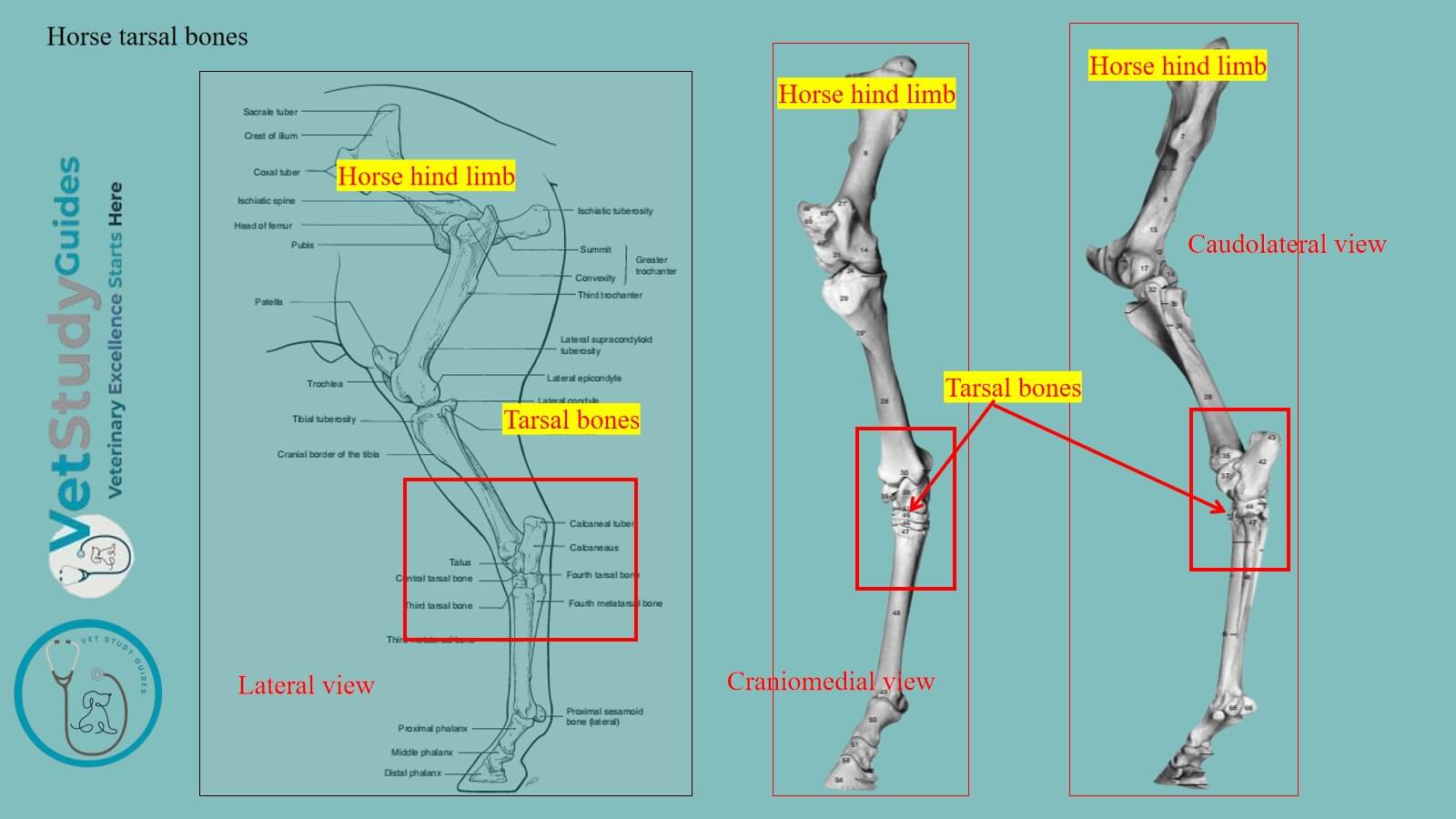
Horse tarsal bones from the tarsus part of the pes segment. In this guide, I will describe the osteological features of the tarsal bones from the horse’s hind limb.
Horse tarsal bones
Where are the tarsal bones in a horse? Location: The tarsal bones in a horse are located between the tibia and fibula bones and the metatarsal bone. They are also known as the tarsus or hock of the horse.
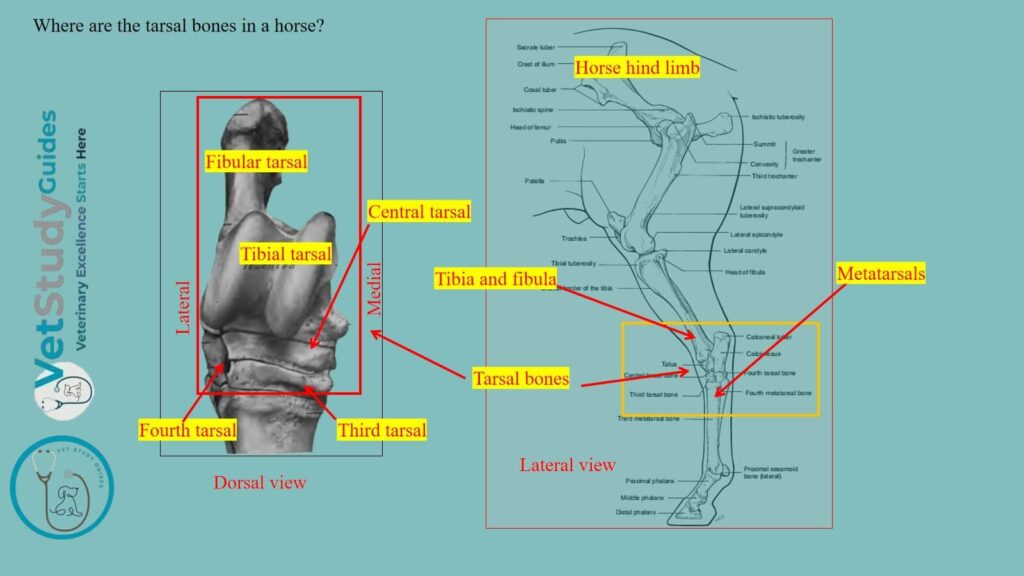
Number of the tarsal bones: Here, the tarsus of a horse comprises six short bones, but exceptionally, seven are present. Table 1 shows the tarsal bones from the horse’s hock –
| Horse tarsals | Name of the tarsals |
| Proximal row | Tibial tarsal (medial), fibular tarsal (lateral) |
| Middle row | Central tarsal |
| Distal row | First and second fused, third tarsal, and fourth tarsal (medial to lateral) |
Thus, the tarsals of the horse are arranged into three rows: proximal, middle, and distal. Here, the proximal row contains tibial and fibular tarsals, and the medial row contains only the central tarsal. However, the distal row contains 3 tarsals: the first and second are fused, the third and fourth tarsals.
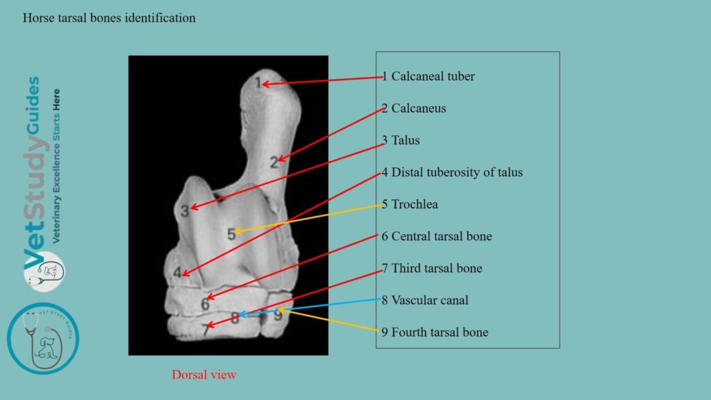
Now, I will describe the horse’s tarsal bones separately, using the diagram. Here, the figure identifies all the tarsals from the horse’s hock/tarsus.
Tibial tarsal bone in a horse
The tibial tarsal bone/os tarsi tibiale is the medial bone of the proximal row. It is extremely irregular in form. However, it may be considered as offering six surfaces for description.
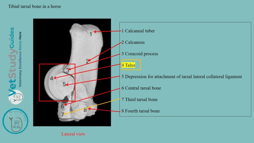
Proximal and distal surfaces: The proximal and dorsal surfaces are continuous and form a trochlea for articulation with the distal end of the tibia.
Trochlea: Here, the trochlea consists of two oblique ridges with a deep groove between them. However, these curves spiral forward, downward, and outward, forming an angle of 12 to 15 degrees with a sagittal plane. There is usually a shallow synovial fossa in the groove.
Distal surface: The distal surface is convex from before backward, and most of it articulates with the central tarsal. However, laterally, it has an oblique facet for the fourth tarsal, and a non-articular groove extends into its surface to its middle.
Plantar surface: The plantar surface is oblique and extremely irregular. It presents four facets for articulation with the fibular tarsal bone.
However, the facets are separated by rough excavated areas, and the largest fossa forms with a corresponding one on the fibular tarsal a cavity termed the sinus tarsi.
Medial surface: The medial surface bears on its distal part a large tuberosity. On its proximal part, there is a small one for the attachment of the medial ligament of the hock joint.
Lateral surface: The lateral surface is smaller than the medial. It is marked by a wide, rough fossa in which the lateral ligament is attached.
Fibular tarsal bone in a horse
The fibular tarsal bone/os tarsi fibulare is the largest bone of the hock. It is elongated, flattened from side to side, and forms a lever for the muscles that extend the hock joint.
Description: It consists of a body and a medial process, the sustentaculum tali.
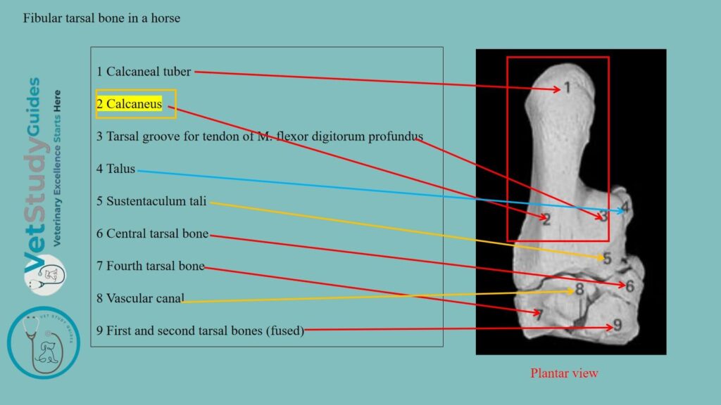
Body of the fibular tarsal
The body is enlarged at its proximal end to form the tuber calcis or “point of the hock”. Here, the posterior part of this eminence gives attachment to the tendon of the gastrocnemius.
However, in front and on each side, the body provides insertions for the tendons of the superficial digital flexor, biceps, and semitendinosus muscles.
Distal extremity: The distal extremity bears a concave facet for articulation with the fourth tarsal bone.
Medial surface: The medial surface of the body has, on its lower part, a strong process. It is known as the sustentaculum tali, which projects inward.
Process: The process has a large, oval, slightly concave facet in front for articulation with the tibial tarsal. It sometimes presents a small articular surface below for the central bone.
Plantar surface: It forms, with the smooth medial surface of the body, a groove for the deep flexor tendon.
Medial surface: Its medial surface has a prominence on the distal part for the attachment of the medial ligament.
Lateral surface: The lateral surface of the body is flattened, except below. There is a rough prominence for the attachment of the lateral ligament below.
Borders of the fibular tarsal bone
Dorsal border: The dorsal border is concave in its length, smooth and rounded in its upper part. About its middle is a blunt and pointed projection.
It bears facets on its medial and lower surfaces for articulation with the tibial tarsal bone. However, it is roughened laterally for ligamentous attachment.
Below this are two facets for the tibial tarsal and an extensive rough fossa, which contributes to the formation of the sinus tarsi.
Plantar border: The plantar border is straight and broad, and widens a little at each end. However, it is rough and gives attachment to the long plantar ligament.
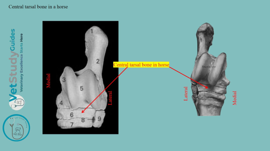
Central tarsal bone in a horse
The central tarsal bone/os tarsi centrale is irregularly quadrilateral. It is situated between the tibial tarsal and the third tarsal.
However, it is flattened from above downward, and may be described as having two surfaces and four borders.
Proximal surface: The proximal surface is concave from before backward. However, almost all of it articulates with the tibial tarsal.
There is a non-articular depression that cuts into its lateral part. Sometimes there is a facet on the posterior angle of the fibular tarsal bone.
Distal surface: The distal surface is convex and is crossed by a non-articular groove. This groove separates facets for articulation with the third and the first and second (fused) tarsals.
Dorsal border: The dorsal and medial borders are continuous, convex, and rough.
Plantar border: The plantar border bears two prominences, separated by a notch.
Lateral border: The lateral border is oblique and bears anterior and posterior facets. They are designed for articulation with the fourth tarsal, between which it is excavated and rough.
First and second tarsal bones
The first and second tarsal bones/os tarsale primum et secundum are usually fused in the horse. Thus, they form one of the very irregularly shaped bones.
Location: These bones are situated in the medio-plantar part of the distal row, below the central and behind the third tarsal.
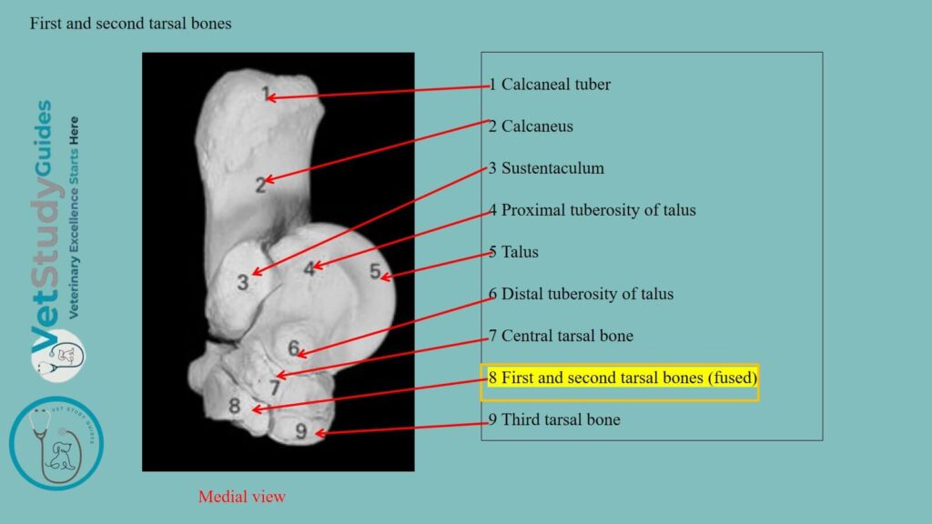
Description: It is the smallest of the tarsal bones, and may be described as having four surfaces and two extremities.
Medial surface: The medial surface faces backward and inward, and is convex. Its anterior part is ridged and gives attachment to the medial ligament.
Again, its posterior part bears an imprint where the medial tendon of the tibialis anterior is inserted.
Lateral surface: The lateral surface is marked by a deep notch, which indicates the division between the first and second tarsal elements. However, it bears, on its anterior part, a facet for the third tarsal.
Proximal surface: The proximal surface is concave and has two facets for articulation with the central tarsal. However, it is separated from the medial surface by a prominent border.
Distal surface: The distal surface is broad in front, where it articulates with the large and medial small metatarsal bones.
Dorsal and plantar extremities: The dorsal extremity bears a ridge or tubercle. The plantar extremity is a blunt point.
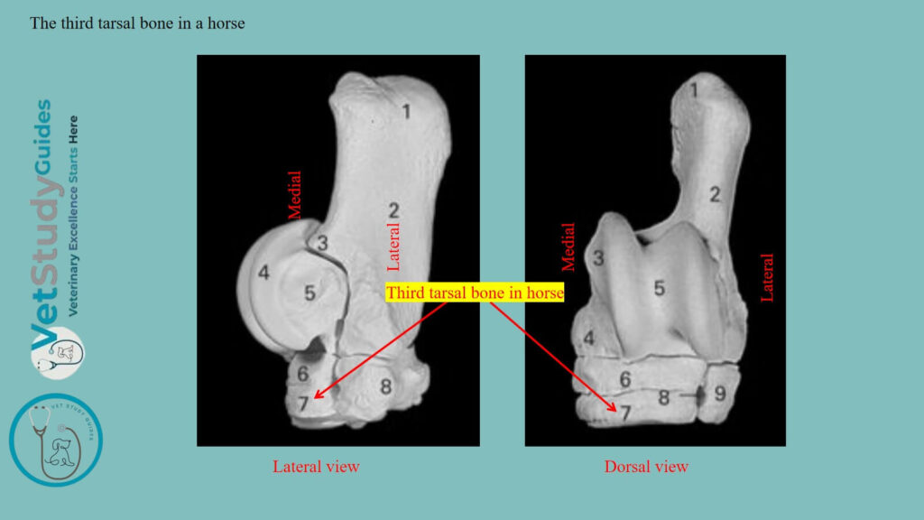
The third tarsal bone in a horse
The third tarsal bone/os tarsale tertium resembles the central, but is smaller and triangular in outline.
Location: It is situated between the central above and the large metatarsal bone below. It possesses two surfaces and three borders.
Proximal surface: The proximal surface is concave and is crossed by a non-articular depression. However, this depression divides it into two unequal facets and articulates with the central tarsal.
Distal surface: The distal surface is slightly convex and rests on the large metatarsal bone. However, it has an extensive central rough excavation.
Dorsal border: The dorsal border is convex and bears a rounded ridge on its medial part.
Medial border: The medial border is deeply notched and has a small facet for the second tarsal on its anterior part.
Lateral border: The lateral border is also divided by a notch into two parts. It bears two diagonally opposite facets for articulation with the fourth tarsal.
Exception: In some cases, there is a facet for the medial small metatarsal bone.
Fourth tarsal in a horse
The fourth tarsal bone/os tarsale quartum is the lateral bone of the distal row. It is equal in height to the central and third together.
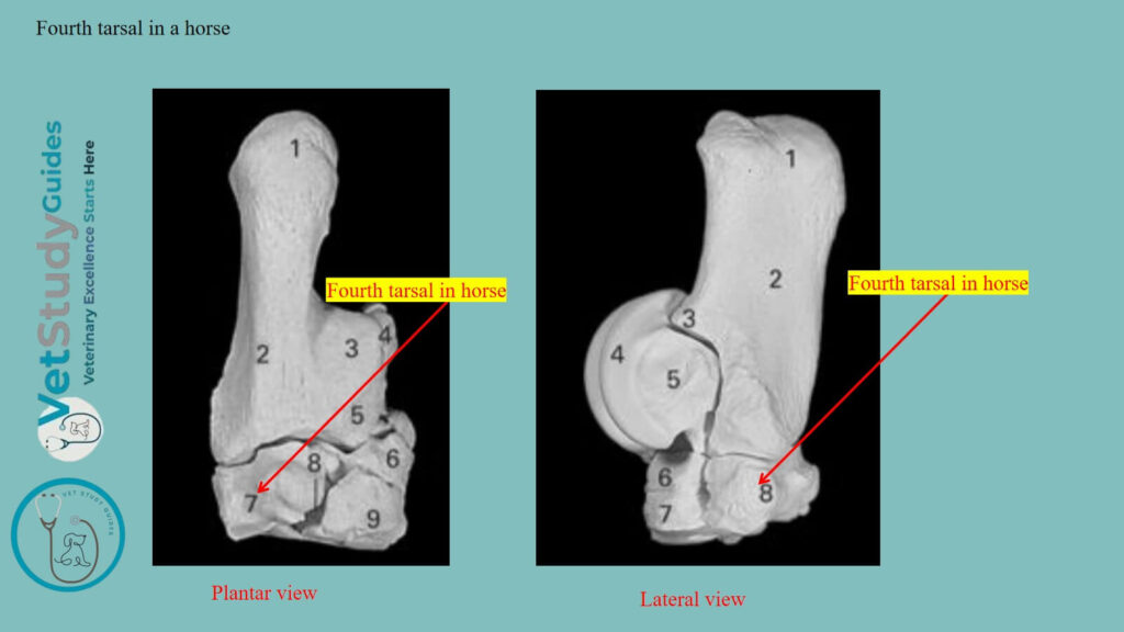
The fourth tarsal of the horse is cuboid in shape and presents six surfaces –
Proximal surface: The proximal surface is convex from side to side and articulates chiefly with the fibular tarsal. However, it also articulates to a small extent with the tibial tarsal.
Distal surface: The distal surface rests on the large and lateral small metatarsal bones.
Medial surface: The medial surface bears four facets for articulation with the central and third tarsal bones. It is crossed by a smooth groove running from the front to the back.
It is apposition with the adjacent bones that forms the canal of the tarsus. However, this canal is designed for the passage of the perforating tarsal vessels.
Dorsal, lateral, and plantar surfaces: The dorsal, lateral, and plantar surfaces are continuous and rough. A tuberosity behind gives attachment to the plantar ligament.
FAQ’s on horses’ tarsal bones
The fibular tarsal bone has two centers of ossification, one for the main mass and the other for the tuber calcis. However, the latter fuses with the rest of the bone at about three years of age. The first and second tarsals have separate centers, but fusion usually occurs before birth. Each of the other bones ossifies from a single center.
Yes, the horse may have more than 6 tarsals. Sometimes they may have 7 tarsals in their hock or tarsus.
In these cases, the first and second tarsal bones remain separate—a remarkable reversion to the condition in the early ancestors of the horse.
In such bones, the first tarsal is a discoid bone, articulating above with the central, below with the small metacarpal bone.
Conclusion
Thus, the horse tarsal bones form the tarsus, and they are arranged into three rows. Here, the first/proximal row consists of the tibial and fibular tarsal, which are facing medially and laterally, respectively.
However, the variation in the number of tarsals in the distal row varies from 6 to 7 bones.
References
- Sisson, S., Anatomy of the domestic animals. W B Saunders Company, USA.
- Dyce and Wensing, Textbook of Veterinary Anatomy, 4th edition, Saunders, USA.
- C. J. Ley, et al. Osteochondral lesions in distal tarsal joints of Icelandic horses reveal strong associations between hyaline and calcified cartilage abnormalities. Eur. Cell. Mater. 2014, 27, 213-236.
- Beth V, and Robert W. C, Veterinary Assisting: Fundamentals & Applications, ISBN-13: 978-1-4354-5387-6, Maxwell Drive, Clifton Park, NY 12065-2919 USA.
- Vladova, D., et al. (2026). A Case of Separate os tarsale primum in a Horse. Veterinary Sciences, 13(6), 582.
- Anna Dee Fails and Christianne Magee, Anatomy and physiology of the Farm Animals, 111 River Street, Hoboken, NJ 07030, USA.
- Hilary M. Clayton, Peter F. Flood, Diana S. Rosenstein, and David Mandeville, Clinical Anatomy of the Horse, First edition 2005, ISBN 07234 3302 X.
- Din, S. et al., Osteometric and Radiographic Studies of Tarsal Bones in Adult Chinkara (Gazella bennettii). Pak journal of zoology, 2019; Vol. 51, Iss. 6, Pages 1999-2399
- Olstad K, et al., Osteochondrosis in the central and third tarsal bones of young horses. Vet Pathol. 2024 Jan;61(1):74-87.
- Pasquini and Spurgeon, Anatomy of domestic animals, systemic and regional approaches.
- Victoria Aspinall B, and Melanie Cappello, Introduction to Veterinary Anatomy and Physiology Textbook, ISBN 978-0-7020-5735-9, Elsevier.
Reviewed and Edited by