The horse tibia and fibula are the bones of the appendicular skeleton. Here, the horse’s tibia is a typical long bone, while the fibula is a reduced long bone of the hindlimb.
Here, you will learn the details features of the horse’s tibia and fibula bones.
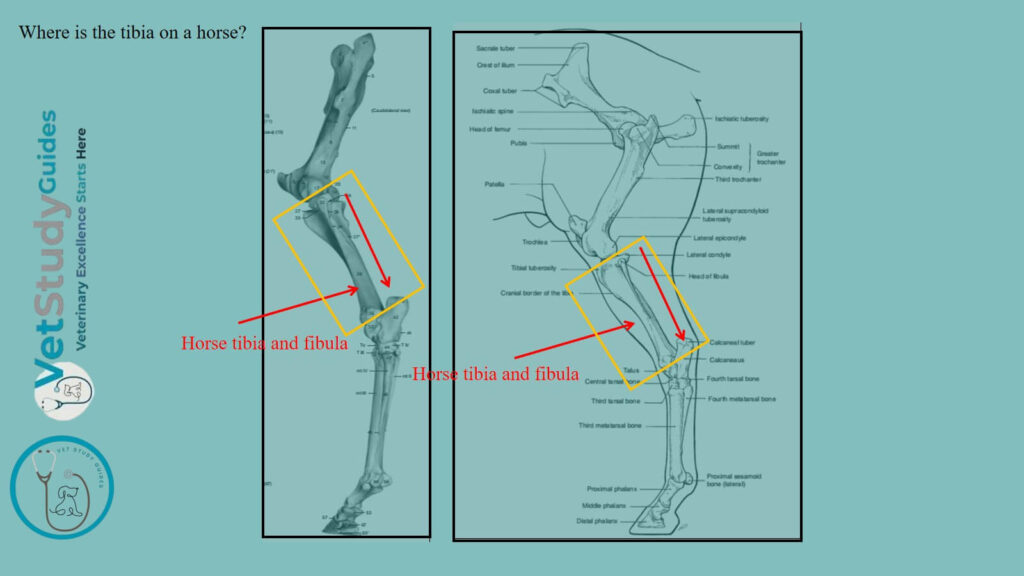
Where is the tibia on a horse?
The horse tibia is a long bone which extends obliquely downward/below and backward/caudal from the stifle to the hock. It articulates above with the femur, below with the tarsus, and laterally with the fibula.
Horse tibia anatomy
For a description of the horse tibia anatomy, you may follow the typical features of a long bone. It possesses a three-sided shaft and two extremities: proximal and distal.
The shaft of the horse tibia
The shaft/corpus tibiae is large and three-sided above. It becomes smaller and flattened in the sagittal direction below.
However, it widens at the distal end. It presents three surfaces (lateral, medial, and posterior) and three borders (anterior, lateral, and medial).
Surfaces of the shaft of the horse tibia
Medial surface: The medial surface/facies medialis is inroad above. Here, it presents rough prominence for the attachment of the medial ligament and the sartorius and gracilis muscles. Again, below this, it is narrower, convex from edge to edge and subcutaneous.
Lateral surface: The lateral surface/facies lateralis is smooth and somewhat spiral. It is wide and concave in its proximal fourth.
Here, it becomes narrower and convex, and winds gradually to the front of the bone. However, near the distal end, it widens a little, becomes flat, and faces forward.
Posterior surface: The posterior surface/facies caudalis is flattened. It is divided into two parts by the rough popliteal line/linea poplitea.
It runs obliquely from the proximal part of the shaft’s lateral border to the middle of the medial border.
The popliteus muscle occupies the triangular area above the line. However, the area below is marked by rough lines/linese musculares.
Here, the deep flexor muscle of the digit is attached. The popliteal lines fade out distally, where the surface is smooth and flat.
Nutrient foramen: The nutrient foramen is situated on or near the popliteal line.
Borders of the tibial shaft
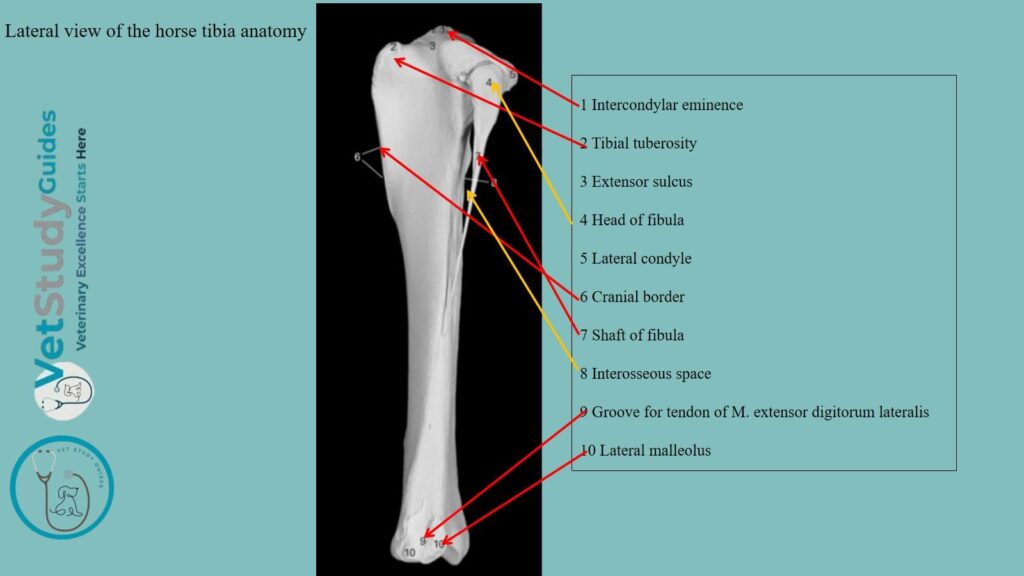
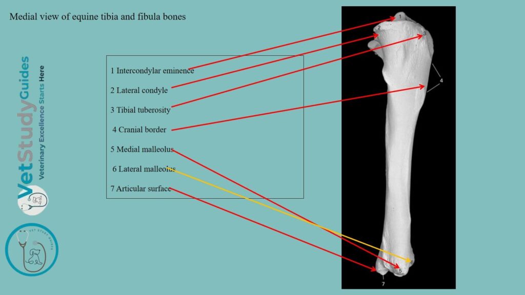
Anterior border: The anterior border is very prominent in its proximal third, forming the crest of the tibia/crista tibiae. Distally, it is reduced to a rough line, which ends at a small elevation near the distal end of the bone.
The medial surface of the crest presents a rough prominence for the attachment of the tendon of the semitendinosus.
Medial border: The medial border/margo medialis is rounded in its proximal half. Here, the popliteus muscle is attached, and a tubercle is found on this part. The distal part is a rough line on well-marked bones.
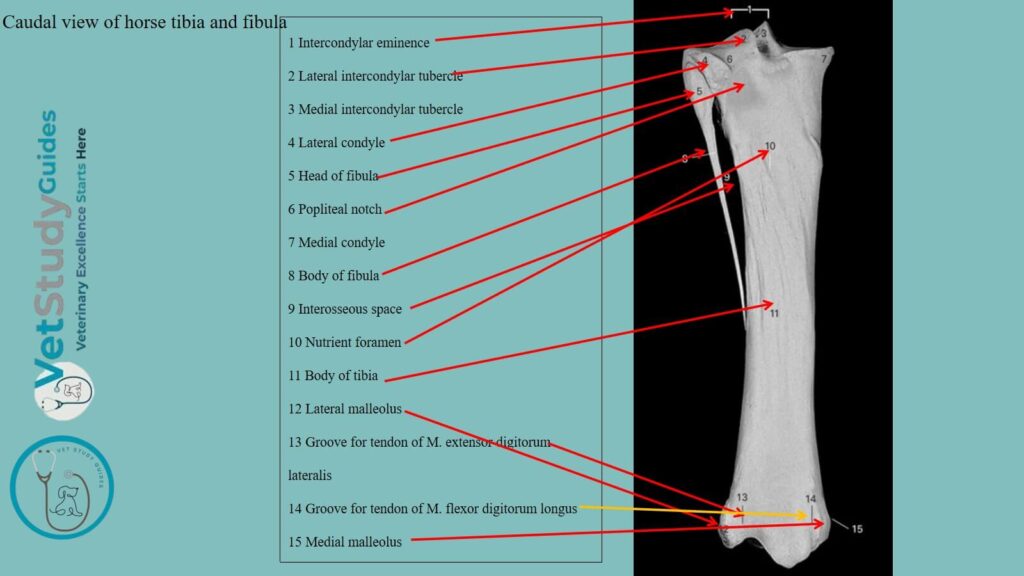
Lateral border: The lateral border/crista interossea is concave in its proximal part. It concurs with the fibula in the formation of the interosseous space of the leg/spatium interosseum cruris.
A smooth impression indicates the course of the anterior tibial vessels through the space to the front of the leg. About the middle of the bone, the border divides and encloses a narrow triangular surface.
Proximal extremity of the horse femur bone
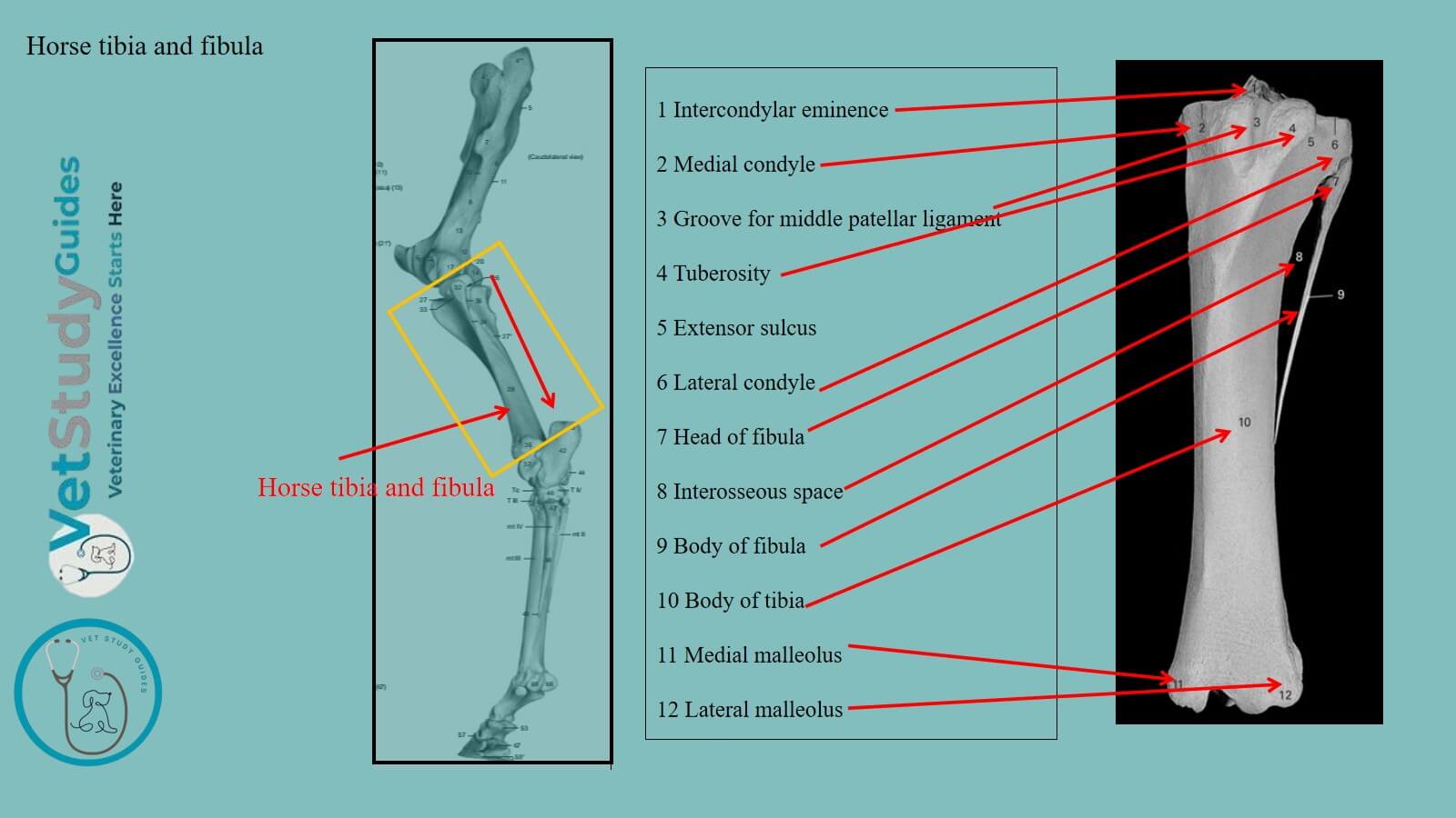
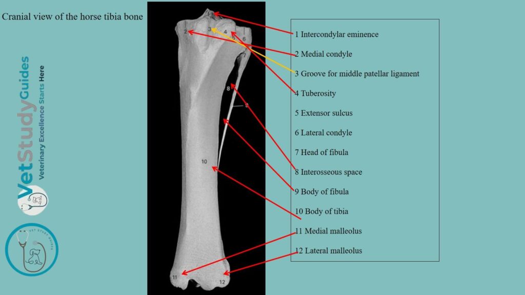
The proximal extremity/extremitas proximalis is large and three-sided. It bears two articular eminences, the medial and lateral condyles/condylus medialis, lateralis.
Articulation: Each presents a somewhat saddle-shaped surface for articulation with the corresponding condyle of the femur and meniscus.
Spine: The spine or intercondyloid eminence is the central prominence. Upon the sides of the spine, the articular surfaces are continued. It consists of a high medial part and a lower lateral part.
On, before and behind the spine are the intercondyloid fossae. Within it, the anterior cruciate ligament and the menisci are attached.
Condyles: The condyles are separated by the deep popliteal notch. On the medial side of this notch, there is a tubercle for the attachment of the posterior cruciate ligament.
Lateral condyle: The lateral condyle has an overhanging outer margin. There is a facet for articulation with the fibula.
The large anterior eminence is the tuberosity of the tibia/tuberositas tibiae. It is marked in front by a groove/sulcus ligamenti.
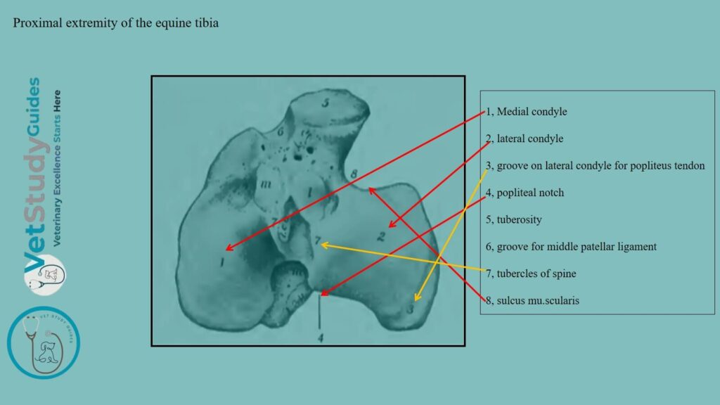
However, the lower part of the tuberosity gives attachment to the middle patellar ligament/ Again, the groove is flanked by rough areas for the attachment of the medial and lateral patellar ligaments.
Notch: A semicircular smooth notch, the sulcus muscularis, separates the tuberosity from the lateral condyle. It gives passage to the common tendon of origin of the extensor digitalis longus and the peroneus tertius.
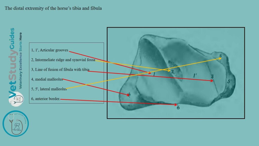
Distal extremity of the horse tibia
The distal extremity/extremitas distalis is much smaller than the proximal one. This extremity is quadrangular in form and larger medially than laterally.
It presents an articular surface, which is adapted to the trochlea of the tibial tarsal bone.
Ridge and grooves: The distal extremity consists of two grooves separated by a ridge. Here, the ridge and grooves are directed obliquely forward and laterally.
They are bound on either side by the malleoli, to which the collateral ligaments of the hock joint are attached. A shallow synovial fossa is usually present in the middle of the articular ridge.
Lateral groove: The lateral groove is wider and shallower than the medial one. It is frequently marked by a line or groove which indicates the former demarcation between the tibia and fibula.
Medial malleolus: The medial malleolus/malleolus medialis is the more prominent of the two. It forms the anterior boundary of a groove for the tendon of the flexor digitalis longus.
Lateral malleolus: The lateral malleolus/malleolus lateralis is broader. A vertical groove marks it for the passage of the lateral extensor tendon.
Horse fibula
Location: The fibula of the horse is a much-reduced long bone, situated along the lateral border of the tibia.
The shaft of the horse fibula
The shaft/corpus fibula is a slender rod which forms the lateral boundary of the interosseous space of the leg. It usually terminates below in a pointed end about one-half to two-thirds of the way down the lateral border of the tibia.
Extremities of the horse’s fibula bone
Proximal extremity: The proximal extremity or head is relatively large and is flattened transversely. Its medial surface presents a narrow area along the upper border. This structure is for articulation with the lateral condyle of the tibia.
The lateral surface is rough and gives attachment to the lateral ligament of the stifle joint. It has rounded anterior and posterior borders.
Distal extremity: The distal extremity is fused with the tibia, constituting the lateral malleolus.
FAQ on the horse’s tibia and fibula bones
The horse tibia has the usual three chief centers of ossification and supplementary ones for the tuberosity and the lateral malleolus. The latter is really the distal end of the fibula. It is a separate piece at birth, and the line of union is commonly quite evident in the adult in the articular groove.
However, the proximal end unites with the shaft at about three and a half years, and the distal end at about two years of age.
The development of the horse fibula bone resembles that of the ulna. The embryonic cartilaginous fibula extends the entire length of the leg, but does not articulate with the femur.
The distal part of the shaft is usually reduced to a fibrous band. Three centers of ossification appear, one each for the shaft and the extremities.
Here, the distal end unites early with the tibia, forming the lateral malleolus. It is interesting to note that in some cases, the entire shaft of the fibula develops. Again, a reversion to the condition in the Miocene ancestors of the horse.
Conclusion
So, the horse tibia and fibula are the long bones of the leg segment of the hind limb. Here, the horse tibia belongs to the typical features of a long bone, where it has a body and two extremities. However, the horse fibula is a reduced long bone that attaches to the lateral aspect of the tibia and possesses a shaft and two extremities.
References
- Sisson, S., Anatomy of the domestic animals. W B Saunders Company, USA.
- Dyce and Wensing, Textbook of Veterinary Anatomy, 4th edition, Saunders, USA.
- Fürst, A., Meier, D., Michel, S., Schmidlin, A., Held, L., & Laib, A. (2008). Effect of age on microarchitecture in the radius and tibia of horses: an Xtreme computed tomographic study. BMC Veterinary Research, 4, 3.
- Anna Dee Fails and Christianne Magee, Anatomy and physiology of the Farm Animals, 111 River Street, Hoboken, NJ 07030, USA.
- Morphometrical studies of tibia, fibula and lateral malleolus of the blue bull (Boselephus tragocamelus), Journal of Entomology and Zoology Studies 2020; SP-8(2): 84-87;
- Hilary M. Clayton, Peter F. Flood, Diana S. Rosenstein, and David Mandeville, Clinical Anatomy of the Horse, First edition 2005, ISBN 07234 3302 X.
- Pasquini and Spurgeon, Anatomy of domestic animals, systemic and regional approaches.
- Devendra Saran, Malvika Maru, Pankaj K Thanvi, Ashok Dangi, Vimala Choudhary, Sandeep Kumar, Raj Kumar Siyag, Prem Chand Tard and Aruna Panwar. Comparative gross anatomical studies on tibia-fibula of Cattle, Horse and Dog. The Pharma Innovation Journal. 2023; 12(7S): 1184-1187.
- Victoria Aspinall B, and Melanie Cappello, Introduction to Veterinary Anatomy and Physiology Textbook, ISBN 978-0-7020-5735-9, Elsevier.