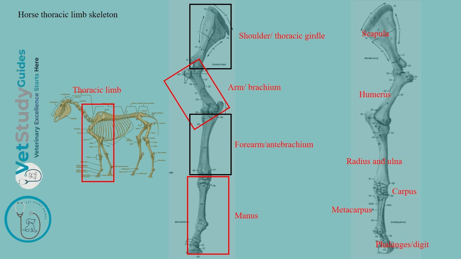
The horse thoracic limb skeleton includes scapula, humerus, radius, ulna, carpals, metacarpals, and phalanges. They are the appendicular part of the horse’s skeleton.
Here, you will learn the overview of the horse’s thoracic limb bones/skeleton. You can learn the details of these bones from dedicated articles.
Horse thoracic limb skeleton
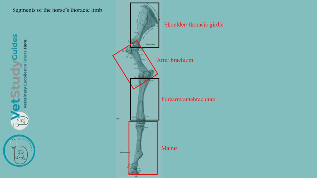
The thoracic limb of a horse consists of four chief segments:
- First segment: The shoulder girdle,
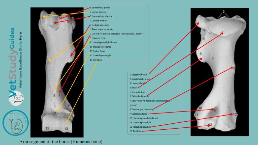
- Second segment: the arm,
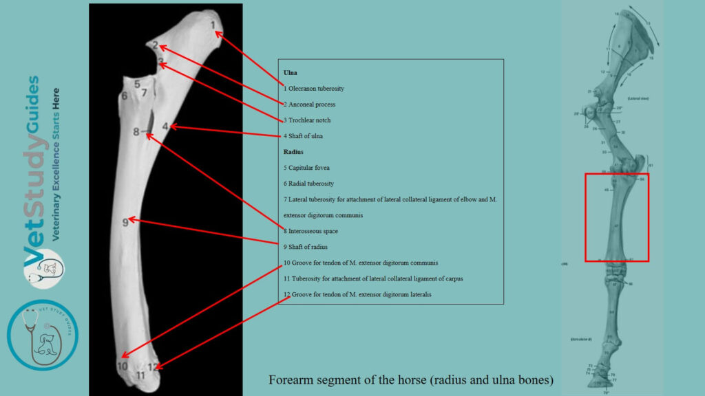
- Third segment: the forearm, and
- Fourth segment: the manus.
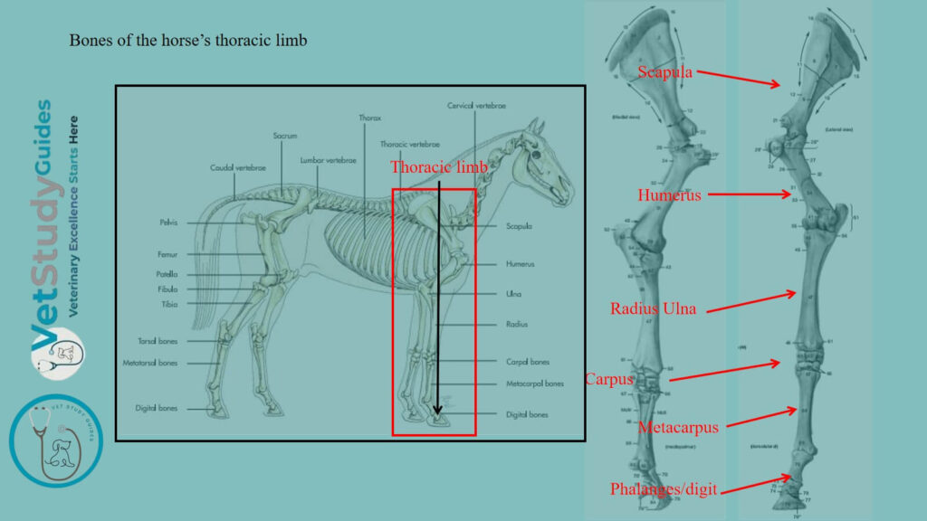
Bones of the horse’s thoracic limb
Thus, you need to know the details of the osteological features of the following bones from the horse’s thoracic limb –
- Scapula (forms the shoulder girdle with coracoid and clavicle),
- Humerus (long bone that forms the arm segment),
- Radius and ulna bones (form the forearm segment),
- Carpal bones of a horse,
- Metacarpal bones of the horse, and
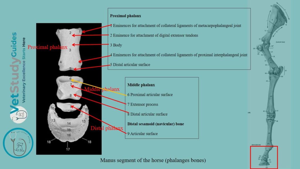
- Phalanges of the horse,
Carpals, metacarpals, and phalanges form the manus segment in the horse’s thoracic limb.
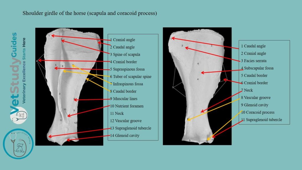
Shoulder girdle of a horse
The shoulder girdle/cingulum extremitatis thoracalis, when fully developed, consists of three bones—
- First, the scapula (or shoulder-blade),
- Second, the coracoid, and
- Third, the clavicle (or collar-bone).
Scapula: Domesticated mammals like the horse have only the scapula. It is a large, flat bone that is well-developed.
A small coracoid element has fused with the horse’s scapula bone. However, the clavicle is either absent or is a small rudiment.
It is embedded in the brachiocephalicus muscle. There is therefore no articulation of the shoulder with the axial skeleton.
The arm and forearm of the horse
The arm/brachium of a horse contains a single long bone, the humerus/arm bone.
In the forearm/antibrachium are two bones, the radius and ulna.
Radius and ulna: These radius and ulna bones of the horse’s forearm vary in relative size and mobility.
Horse and ox: In the horse and ox, the two bones are fused. Again, the distal part of the limb is fixed in the position of pronation.
The radius is placed in front and supports the weight. The ulna is well developed only in its proximal part. It forms a lever for the extensor muscles of the elbow.
Pig: In the pig, the ulna is the larger and longer of the two bones. But it is closely attached to the back of the radius.
Dog: In the dog, the ulna is also well developed, and a small amount of movement is possible between the two bones.
Manus of the horse
The manus is the homologue of the hand in man. It consists of three subdivisions –
- First, the carpus,
- Second, metacarpus, and
- Third, digit or digits.
Carpus of the horse
Carpus: The carpus, popularly termed the knee in animals. It is homologous with the wrist of a man.
Here, the carpus of the horse contains a group of short bones, the ossa carpi. These are typically eight in number and are arranged in two transverse rows –
- A proximal or antibrachial, and
- A distal or metacarpal.
Proximal row carpals: The bones of the proximal row, named from the radial to the ulnar side (from within outward), are the radial, intermediate, ulnar, and accessory carpal bones.
Distal row carpals: The bones of the distal row are designated numerically, in the same direction, as first, second, third, and fourth carpal bones.
The central carpal bone/os carpi centrale is omitted in the horse carpus since it is not a separate element in the animals under consideration here.
However, Table 1 shows the names and symbols of the horse’s carpal bones –
| Name | Anatomical name | Symbol |
| Radial/scaphoid | Os carpi radiale | Cr |
| Intermediate /semilunar | Os carpi intermedium | Ci |
| Ulnar/cuneiform | Os carpi ulnare | Cu |
| Accessory/pisiform | Os carpi accessorium | Ca |
| First carpal /trapezium | Os carpale primum | C1 |
| Second carpal/trapezoid | Os carpale secundum | C2 |
| Third carpal/os magnum | Os carpale tertium | C3 |
| Fourth carpal/unciform | Os carpale quartum | C4 |
Metacarpal bones of the horse
General information: The metacarpus of an animal typically contains five metacarpal bones/ossa metacarpalia I-V), one for each digit. They are long bones and are designated numerically from the radial to the ulnar side (i. e., from within outward).
This arrangement occurs in the dog, although the first metacarpal is much smaller than the others. The second and fifth are somewhat reduced.
Further reduction has taken place in the other animals, resulting in the artiodactyls and pterodactyl forms.
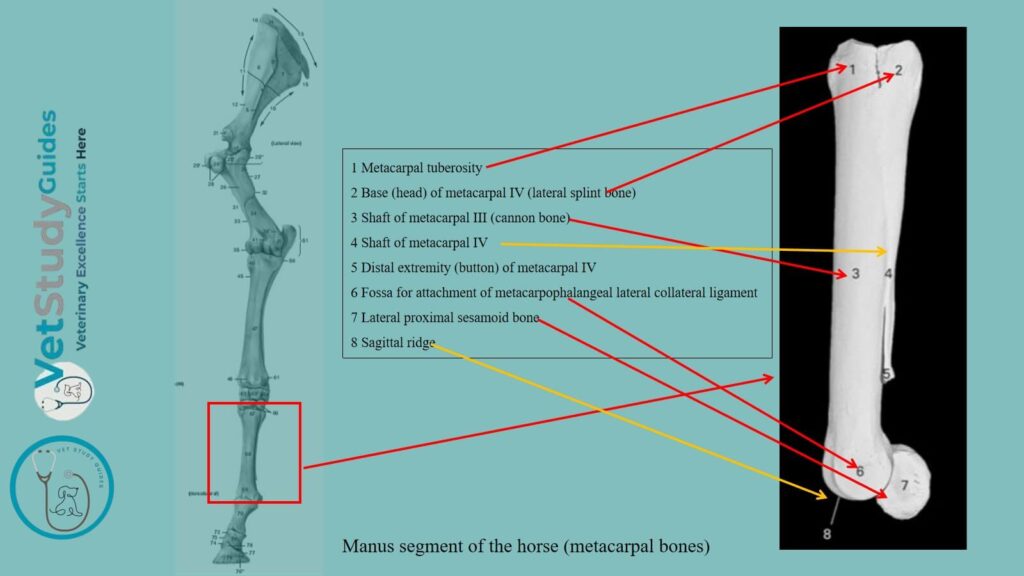
Horse: In the horse, the first and fifth metacarpals are absent, the third is the large supporting metacarpal bone and carries the single digit. Again, the second and fourth are much reduced.
Other animals: In artiodactyls (ox, sheep, pig), the third and fourth are the chief metacarpals and carry the well-developed digits. They are fused in the ox and sheep.
The others are variously reduced or absent, as noted in the special descriptions to follow. The fossil remains of the ancestors of the existing equids illustrate most completely the reduction which has occurred in this respect(Getty).
The first metacarpal bone was small. Intermediate forms show the gradual evolution of the race from this primitive animal, which was about the size of the domestic cat.
Digits in horses and other animals
General information: There is reason to believe that earlier forms had five digits. The digits/digiti manus are homologous with the fingers of man.
They are typically five in number. They are designated numerically from the radial to the ulnar side, in correspondence with the metacarpus.
Dog, ox, and pig: The full number is present in the dog. In the ox and pig, the third and fourth are well developed. They support the weight, while the second and fifth are reduced.
Horse: The existing horse has a single digit, the third of its pentadactyl ancestors. The skeleton of each fully developed digit consists of three phalanges and certain sesamoid bones.
Phalanges and sesamoid bones in a horse
Phalanges: The first phalanx/phalanx prima articulates with the corresponding metacarpal bone above. It also articulates with the second phalanx/phalanx secunda below.
The third phalanx/phalanx tertia is enclosed in the hoof or claw. It is modified to conform to the latter.
Sesamoids: The sesamoid bones/ossa sesamoidea are developed along the course of tendons or in the joint capsules. At these points, these sesamoid bones reduce the pressure.
Proximal sesamoids: Two proximal sesamoids/ossa sesamoidea phalangis primse occur at the flexor side of the metacarpo-phalangeal joint. They form a pulley for the flexor tendon.
Distal sesamoids: The distal sesamoid/os sesamoideum phalangis tertise is similarly placed between the deep flexor tendon and the joint between the second and third phalanx. It is absent in the dog, which has a small sesamoid on the extensor side of the metacarpophalangeal joint, and often at the proximal interphalangeal joint also.
FAQ’s on horse thoracic limb skeleton
The shoulder girdle is fully developed in birds and the lower mammals. In the higher mammals, the coracoid is reduced to the coracoid process of the scapula, and the development of the clavicle conforms with the function of the humerus.
Thus, in typical quadrupeds, such as the horse and ox, in which the forelimbs are used only for support and locomotion, the clavicle is absent.
Other animals which use these limbs for grasping, burrowing, and chewing have a well-developed clavicle that connects the scapula with the sternum.
Numerous cases are recorded of the occurrence of supernumerary digits in the horse and other animals. In some pigs, on the other hand, the two chief digits are fused, and the condition appears to be inherited.
Conclusion
Thus, the horse thoracic limb skeleton comprises the scapula, humerus, radius and ulna, carpus, metacarpus, and digits. All these bones form four distinct segments in the horse’s thoracic limb.
You will find a variation in the appearance and also in the number of bones in the horse’s thoracic limb compared to other animals.
References
- Sisson, S., Anatomy of the domestic animals. W B Saunders Company, USA.
- Dyce and Wensing, Textbook of Veterinary Anatomy, 4th edition, Saunders, USA.
- Ahrari-Khafi, M. S., A. Tabatabaei Naeini & N. Ajvadi, 2018. Ultrasonographic evaluation of normal scapula in the horse. Bulg. J. Vet. Med., 21, No 1, 50–58.
- Anna Dee Fails and Christianne Magee, Anatomy and physiology of the Farm Animals, 111 River Street, Hoboken, NJ 07030, USA.
- de Alcântara Leite dos Reis, D., Gouveia, B.L.R., Júnior, J.C.R. et al. Comparative evaluation of anatomical details of thoracic limb bones of a horse to those of models produced via scanning and 3D printing. 3D Print Med 5, 13 (2019). https://doi.org/10.1186/s41205-019-0050-2
- Solounias, N., Danowitz, M., Stachtiaris, E., Khurana, A., Araim, M., Sayegh, M., & Natale, J. (2018). Evolutionary anatomy of the horse’s manus. Royal Society Open Science, 5(1), 171782. https://doi.org/10.1098/rsos.171782
- Hilary M. Clayton, Peter F. Flood, Diana S. Rosenstein, and David Mandeville, Clinical Anatomy of the Horse, First edition 2005, ISBN 07234 3302 X.
- Brokken, M. and Tucker, R. (2010). The metacarpal/metatarsal region. In Equine MRI, R.C. Murray (Ed.).
- Pasquini and Spurgeon, Anatomy of domestic animals, systemic and regional approaches.
- Gündemir, O., Szara, T., Pazvant, G., Erdikmen, D. O., Duro, S., & Perez, W. (2021). Radiographic Analysis of the Thoracic Limb Phalanges in Arabian Horses and Thoroughbred Horses. Animals, 11(8), 2205.
- Victoria Aspinall B, and Melanie Cappello, Introduction to Veterinary Anatomy and Physiology Textbook, ISBN 978-0-7020-5735-9, Elsevier.