The horse scapula anatomy describes the location and different osteological features of the scapula. It is the ideal flat bone of the forelimb of the horse skeleton.
In this guide, you will learn the details anatomy of the horse scapula bone with diagrams and pictures.
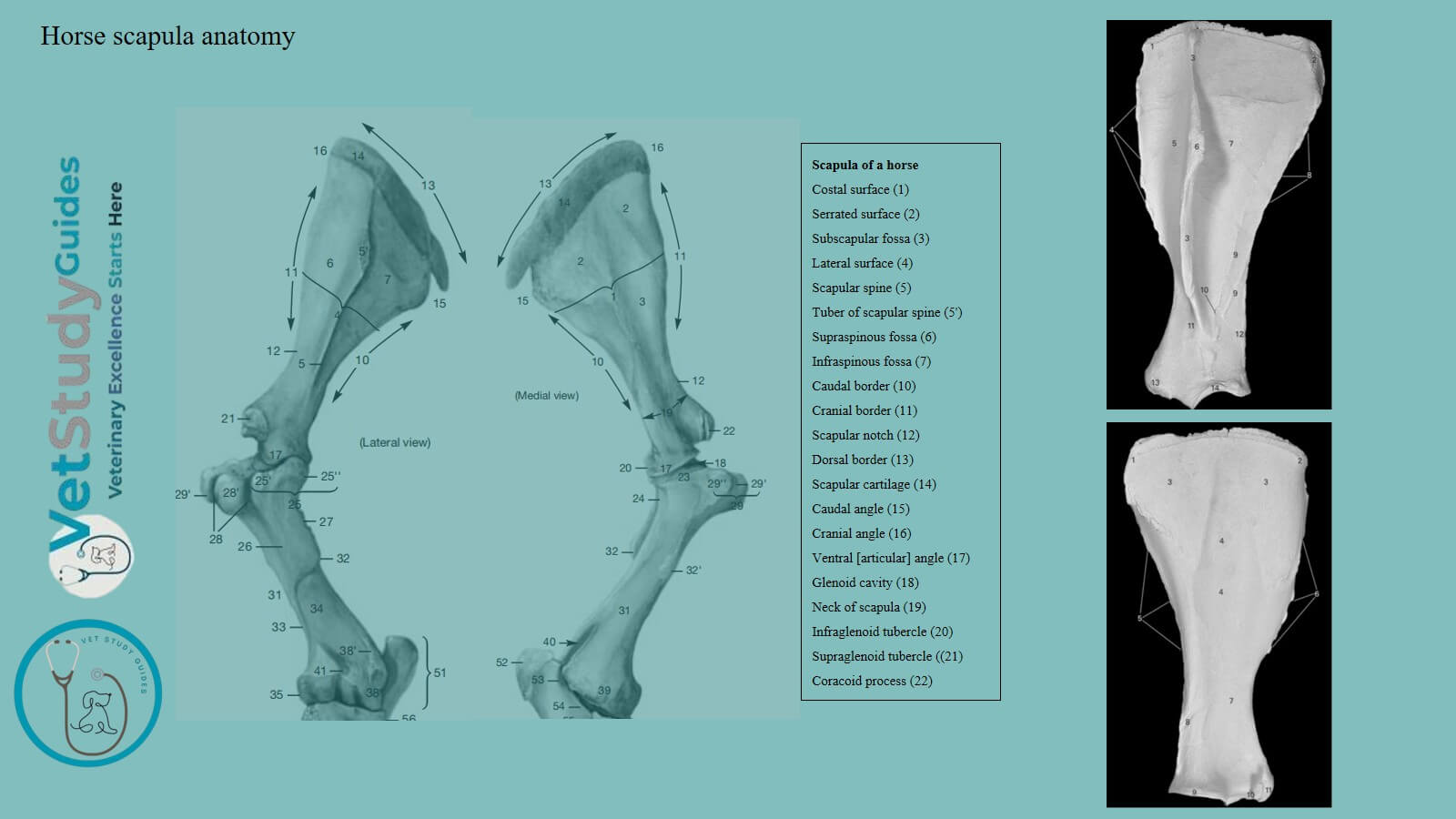
Horse scapula anatomy
You need to describe the followings in a horse scapula bone –
- Location of the horse scapula
- Surfaces of the scapula
- Borders of the scapula
- Angles of the scapula
Location of the horse scapula
The scapula is a flat bone which is situated on the anterior part of the lateral wall of the thorax. Its long axis extends obliquely from the fourth thoracic spine to the sternal end of the first rib.
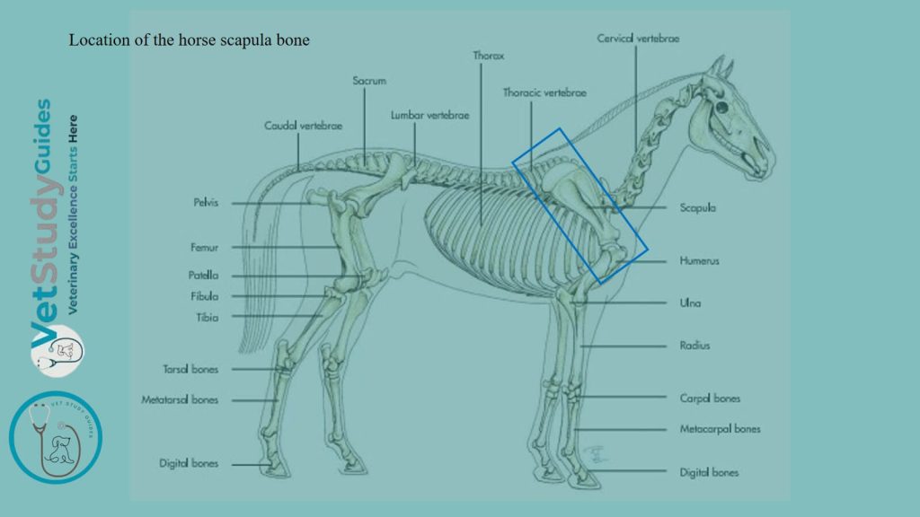
The horse scapula is curved slightly and slopes outward in adaptation to the form of the thoracic wall. It is triangular in outline and has two surfaces, three borders, and three angles.
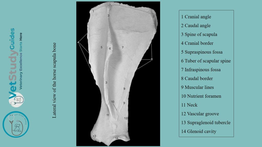
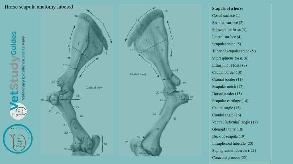
Surfaces of the horse scapula
Lateral surface of the scapula
The lateral surface/facies lateralis is divided into two fossae by the horse’s scapula spine. It extends from the vertebral/dorsal border to the neck of the bone, where it subsides.
The free edge of the spine is thick, rough, and in great part subcutaneous. A little above its middle, there is a variable prominence. This is the tuber spinas/tuberosity of the spine, to which the trapezius muscle is attached.
The supraspinous fossa is situated in front of the spine, and the infraspinous fossa behind it.
The former is much smaller than the latter; it is smooth and is occupied by the supraspinatus muscle. The infraspinous fossa lodges the infraspinatus muscle.
It is wide and smooth in its upper part, narrower below. However, it is marked by several rough lines for muscular attachment. Again, near the neck, there is the nutrient foramen, and a little lower is a vascular groove.
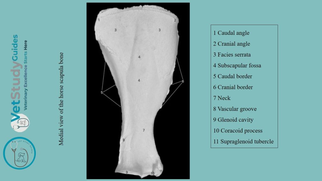
Costal surface of the scapula
The costal surface is hollowed in its length by the subscapular fossa/fossa subscapularis. This fossa occupies nearly the whole of the lower part of the surface.
It is pointed out above and separates two rough triangular areas known as facies serrata. Within this structure, the serratus ventralis is attached. In the lower third, there is a vascular groove with several branches.
Borders of the horse/equine scapula
The anterior border/margo cranialis is convex and rough above, concave and smooth below.
The posterior border/margo caudalis is slightly concave. It is thick and rough in its upper third, thin in its middle, and thickens again below.
The vertebral border/margo vertebralis carries the scapular cartilage.
In the young subject, this edge of the bone is thick and is pitted by impressions into which the cartilage fits. The cartilage is the unossified part of the foetal scapula.
Its lower edge fits the depressions and elevations of the bone. It thins out toward the free edge, which is convex and lies alongside the vertebral spines.
In front, it continues the line of the bone, but behind it forms a rounded projection.
The lower part of the cartilage undergoes more or less ossification. Thus, the vertebral border of the bone in old subjects is thin, irregular, and porous.
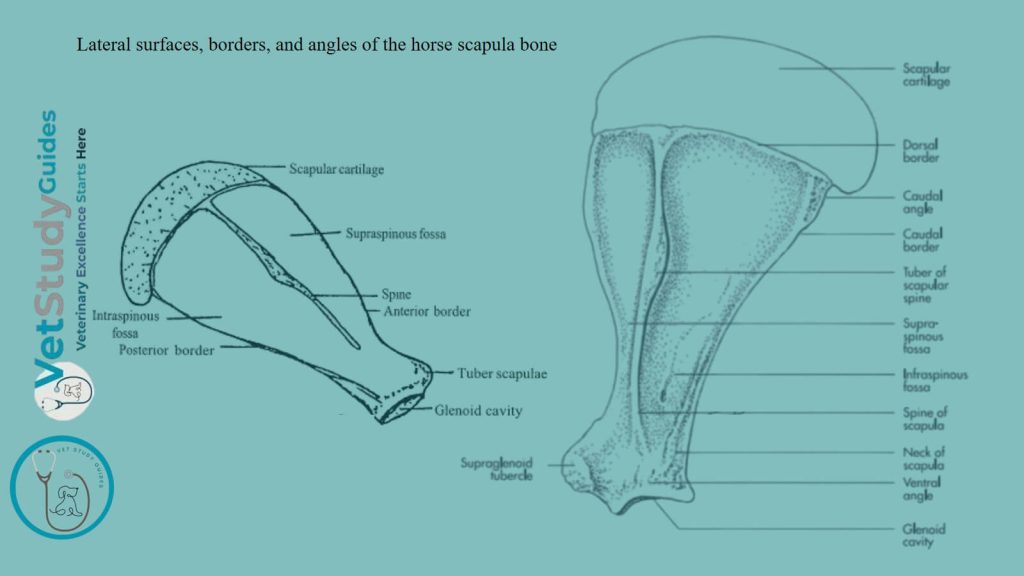
Angles of the horse scapula
Anterior angle of the scapula: The anterior or cervical angle is at the junction of the anterior and vertebral borders and lies opposite to the second thoracic spine. It is relatively thin and is about a right angle.
Posterior angle of the scapula: The posterior or dorsal angle is thick and rough. It is opposite to the vertebral end of the seventh rib. The position can be determined readily in the living animal.
The glenoid angle: The glenoid or articular angle is joined to the body of the bone by the neck of the scapula. It is enlarged, especially in the sagittal direction.
This angle bears the glenoid cavity for articulation with the head of the humerus. The cavity is oval in outline, and its margin is cut into in front by the glenoid notch.
It is also rounded off laterally, just above its postero-lateral part, there is a tubercle. Within this tubercle, a tendon of the teres minor is attached.
The tuber scapulae is the large rough prominence in front. The tendon of origin of the horse’s biceps brachii is attached to the tuber scapulae.
A small coracoid process projects from its medial side, from which the coracobrachialis muscle arises.
Development of the horse scapula bone
The scapula has four centers of ossification, one each for the body of the bone, the tuber scapulae and coracoid process, the anterior part of the glenoid cavity, and the tuber spinae.
The last ossifies after birth and fuses with the spine about the third year. The tuber scapulae and coracoid process fuse with the body of the bone about the end of the first year.
Conclusion
Thus, the horse scapula anatomy describes the location, surfaces, borders, angles, and their unique features.
References
- Sisson, S., Anatomy of the domestic animals. W B Saunders Company, USA.
- Gabriele Barros Mothé, Anselmé Dutra, and Francisco Mendes Junior, Descriptive osteology of the equine thoracic limb. RCMOS – Multidisciplinary Scientific Journal of O Saber. ISSN: 2675-9128. São Paulo-SP, Year IV, v.1, n.1, Jan./Jul. 2024.
- Dyce and Wensing, Textbook of Veterinary Anatomy, 4th edition, Saunders, USA.
- Ahrari-Khafi, M. S., A. Tabatabaei Naeini & N. Ajvadi, 2018. Ultrasonographic evaluation of normal scapula in the horse. Bulg. J. Vet. Med., 21, No 1, 50–58. https://bjvm.trakia-uni.bg/BJVM-March%202018%20p.50-58.pdf
- Beth Vanhorn and Robert W. Clark, Veterinary Assisting: Fundamentals & Applications, ISBN-13: 978-1-4354-5387-6, Maxwell Drive, Clifton Park, NY 12065-2919 USA.
- Anna Dee Fails and Christianne Magee, Anatomy and physiology of the Farm Animals, 111 River Street, Hoboken, NJ 07030, USA.
- Hilary M. Clayton, Peter F. Flood, Diana S. Rosenstein, and David Mandeville, Clinical Anatomy of the Horse, First edition 2005, ISBN 07234 3302 X.
- Pasquini and Spurgeon, Anatomy of domestic animals, systemic and regional approaches.
- Victoria Aspinall B, and Melanie Cappello, Introduction to Veterinary Anatomy and Physiology Textbook, ISBN 978-0-7020-5735-9, Elsevier.