Introduction to veterinary anatomy is concerned with the structure and forms of the organs and tissues of principal domestic animals and birds.
It includes the study of shape, weight, color, texture, and relative position of various organs and tissues of the organisms when they are in a state of normal health.
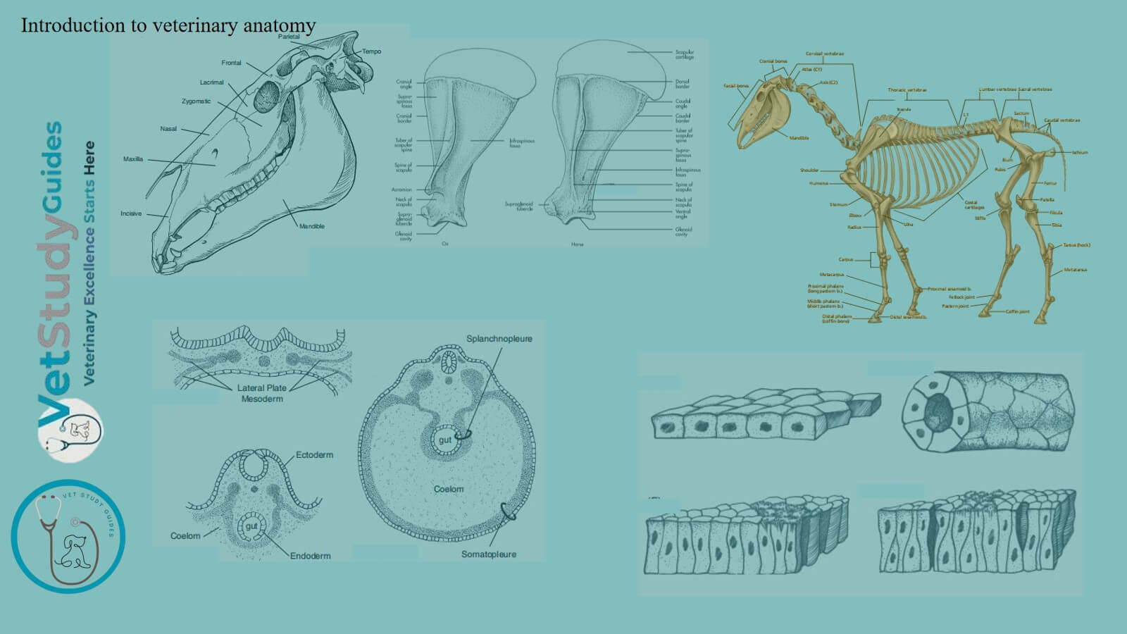
Introduction to veterinary anatomy
Generally veterinary anatomy subject is studied on a comparative basis. However, the biological structures may be studied in different forms.
In gross anatomy, the structures are studied after dissection, with the help of the naked eye only.
Histology or microscopic anatomy deals with the study of the finer details of the structures of various tissues and of different organs. The process of differentiation, growth and development of an organ or structure comes under embryology or developmental anatomy.
When this science of structures and forms of various organ systems of a body is studied in close correlation with the physiology, makes the foundation of the whole of medicine and life science.
Functions of veterinary anatomy
The anatomical knowledge helps a veterinarian, in the discharge of his multifarious duties, in many ways. More important functions of anatomy are:
- It gives an idea about the form, shape, structure and relative position of various organs and tissues comprising a normal and healthy body.
- It aids in understanding the functions of various parts, their correlation and coordination, and their physical and functional relationship.
- When normal shape, structure and function are known, a deviation from this normal can be recognized with great ease. Thus, it helps in the diagnosis and recognition of the diseased state.
- It provides knowledge, which helps in devising the ways and means for the collection of material required for a definite diagnosis.
- It facilitates the development of more efficient means for the administration of drugs, not to mention reducing the drug administration hazards.
- A good grasp of anatomy gives much help and confidence to a surgeon.
Divisions of veterinary anatomy
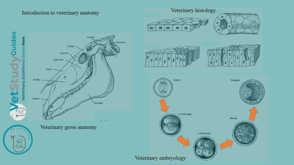
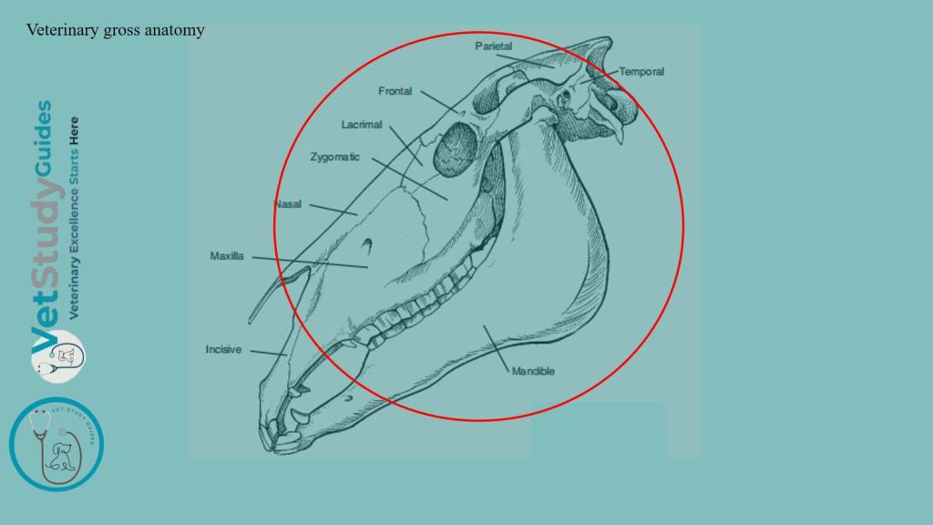
Veterinary gross anatomy: It is the study of forms and structures that can be dissected and observed with the naked eye.
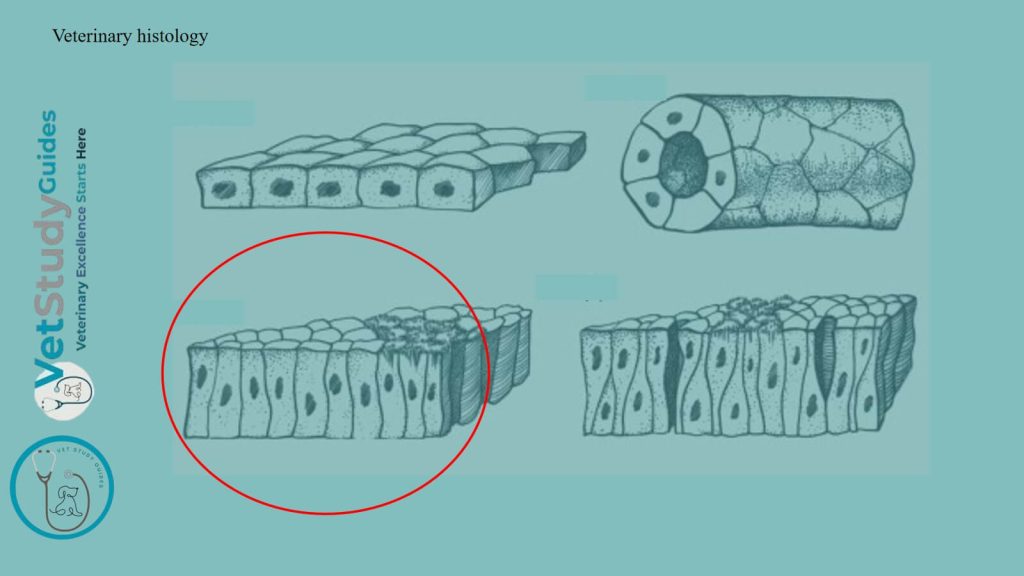
Microscopic anatomy or Veterinary histology: It is the study of minute structures too small to be seen without a microscope.
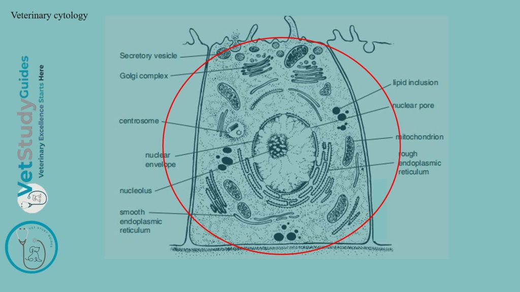
Cytology: It is the study of minute structures in even greater detail, with the help of an electron microscope.
Morbid anatomy: It is the study of diseased structures of an organism.
Applied anatomy: It is the practical application of a normal structural study in relation to the diagnosis and treatment of pathology or a surgical condition. Teratology: It is the study of abnormal development of an individual, including their nature and the causes of the problem.
Developmental anatomy: It is the study of the development of an individual from the zygote to adult.
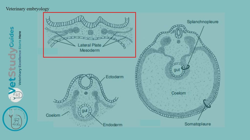
Veterinary embryology: It is the study of the structure from the fertilized egg to birth.
Comparative anatomy: It is the study of the comparison of the structures of animals, and it forms the basis of their classification.
Morphological or philosophical anatomy: It is the study of generalization made on outer form, inner structure, and development of individuals belonging to different species.
Phylogeny: It is the study of the evolutionary history of specie e.g. the study of changes that occurred during the process of evolution.
Special anatomy: It is the description of the structure & form of a single type or specie e.g. anatomy of a horse.
Methods of study in veterinary/animal anatomy
There are two chief methods of study which are usually employed –
- Systemic Study
- Topographic Study
Systemic study of anatomy
In this approach, the body is regarded as consisting of organs or apparatus which are similar in origin and structure. They are also associated with the performance of certain functions.
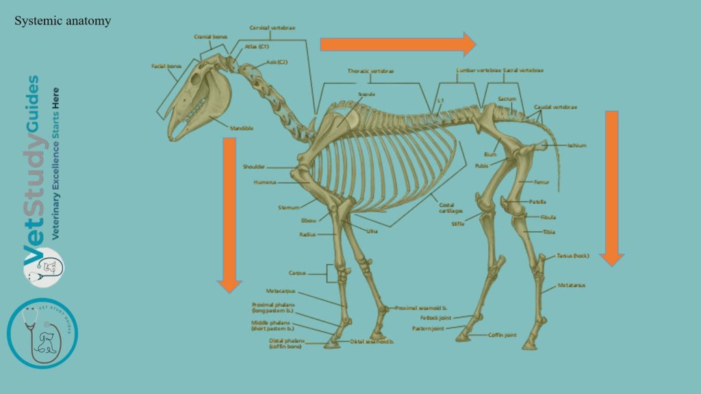
Division of systemic anatomy
- Osteology: The description of the skeleton.
- Syndesmology: The description of the Joints.
- Myology: The description of the muscles.
- Splanchology: The description of the Viscera. This includes the following subdivisions of the soft organs of the body: Digestive System, Respiratory System, Urinary System, genital System (Reproductive System)
- Cardiovascular system: The description of the organs of circulation.
- Nervous system: The description of the nervous system
- Aesthesiology: The description of the sense organs/apparatus and common integument of the animals.
Topographic study method
The term topographic anatomy designates the method/way by which the relative positions of various parts of the animal’s body are accurately determined. Topographic terms are some special terms which are usually employed in anatomical study. It is assumed that the animal is in a standing position.
Other study methods
Topographic Anatomy or Surface Anatomy: In this method, the principal structures or organs of any part of the body are described in relation to a definite and limited area of the surface.
Surgical Anatomy: This is an applied part of the subject where the structures of some specific sites are studied for application in surgical practice.
Radiological Anatomy: Here, the structures of the body are studied with the help of X-rays. Ultrasonography or MRI may be useful for studying the anatomy of any animal in any desired plane. A CT scan is another similar device for studying this subject in normal and altered physiological conditions.
Comparative Anatomy: In this method, analogous structures of different species of animals are studied on a comparative basis.

Recumbency positions
There are the following recoumbency positions –
- Dorsal Recumbency
- Ventral Recumbency
- Lateral Recumbency
Conclusion
So, the introduction to veterinary anatomy provides the basic idea of the anatomy, its classification, functions, and the study methods. Again, it also provides ideas on different terminology that are useful for learning veterinary anatomy.
References
- Sisson, S., Anatomy of the domestic animals. W B Saunders Company, USA.
- Gabriele Barros Mothé, Anselmé Dutra, and Francisco Mendes Junior,
- Dyce and Wensing, Textbook of Veterinary Anatomy, 4th edition, Saunders, USA.
- Anna Dee Fails and Christianne Magee, Anatomy and physiology of the Farm Animals, 111 River Street, Hoboken, NJ 07030, USA.
- Hilary M. Clayton, Peter F. Flood, Diana S. Rosenstein, and David Mandeville, Clinical Anatomy of the Horse, First edition 2005, ISBN 07234 3302 X.
- Pasquini and Spurgeon, Anatomy of domestic animals, systemic and regional approaches.
- Victoria Aspinall B, and Melanie Cappello, Introduction to Veterinary Anatomy and Physiology Textbook, ISBN 978-0-7020-5735-9, Elsevier.