The horse humerus bone is the long type of bone of its forelimb’s skeleton. This guide will help you learn the details anatomy of the horse’s humerus bone.
Location and direction of the horse’s humerus bone
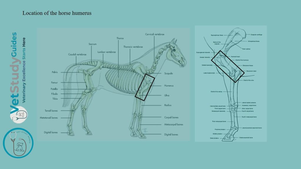
Location: The horse humerus extends from the shoulder above to the elbow below and behind. Above, it articulates with the scapula. However, below it articulates with the radius and ulna bones.
Direction: This bone is directed obliquely downward and backward. It forms an angle of about 55 degrees with the horizontal plane of the horse’s body.
Description of equine/horse humerus bone
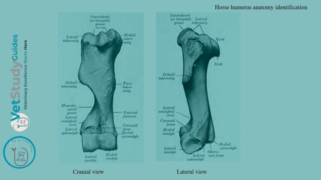
The humerus of a horse consists of a shaft and two extremities (proximal and distal). The shaft is irregularly cylindrical and has a twisted appearance. It may be regarded as having four surfaces: lateral, medial, anterior, and posterior.
Surface of the horse’s humerus bone
Lateral surface of the body/shaft
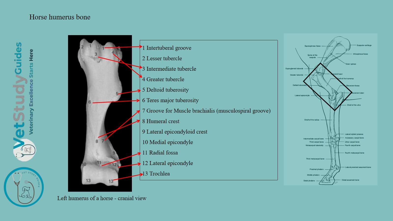
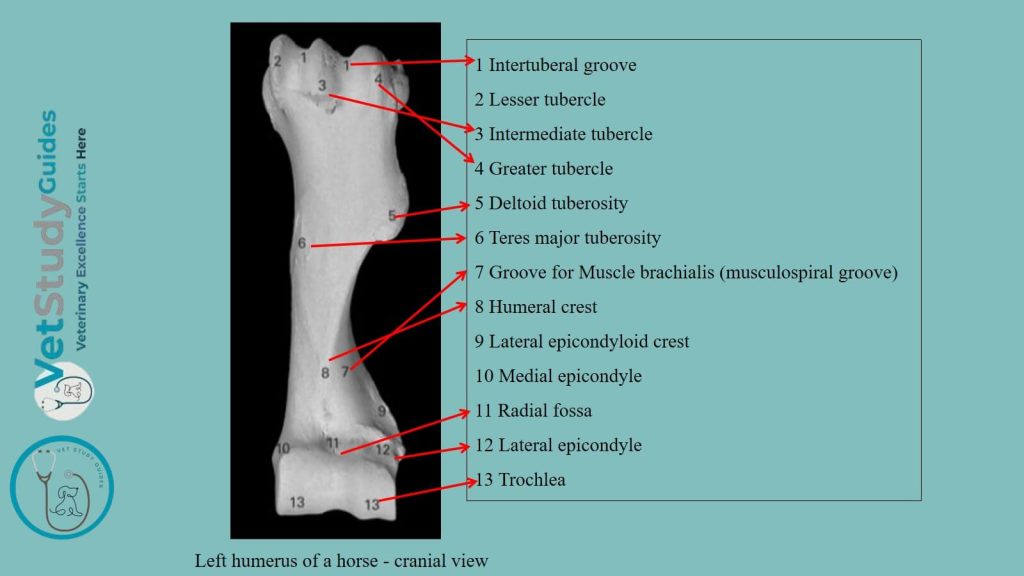
The lateral surface of the horse’s humerus is smooth and is spirally curved. It forms the musculospiral groove, which contains the brachialis muscle.
However, the groove is continuous with the posterior surface above and winds around toward the front below.
Medial surface of the body
The medial surface is nearly straight in its length, rounded from side to side. It blends with the anterior and posterior surfaces.
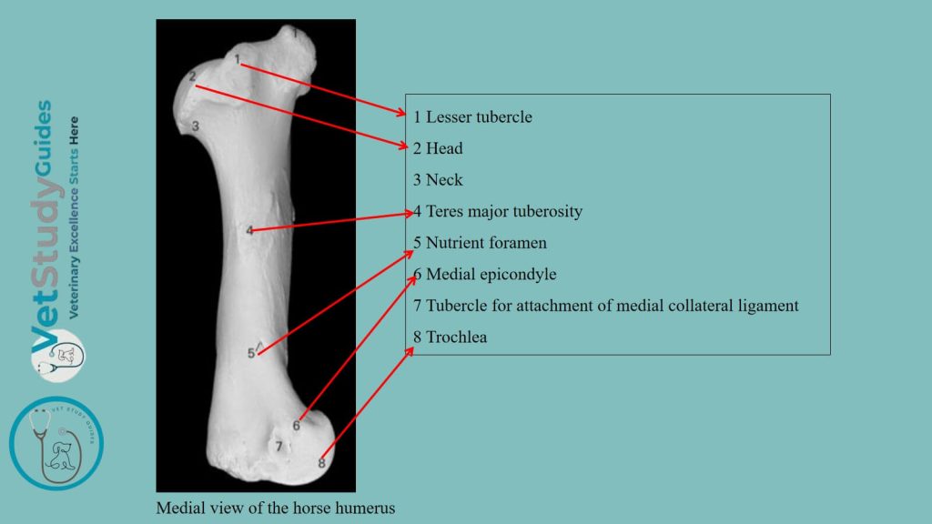
Just above its middle is the teres tuberosity/tuberositas teres. Within this structure, the tendons of the latissimus dorsi and teres major muscles are attached. The nutrient foramen is in the distal third of this surface.
Anterior and posterior surfaces of the humerus
The anterior surface/facies cranialis is triangular, wide and smooth above. Again, this surface is narrow and roughened below.
It is separated from the lateral surface by a distinct border, the crest of the humerus/crista humeri.
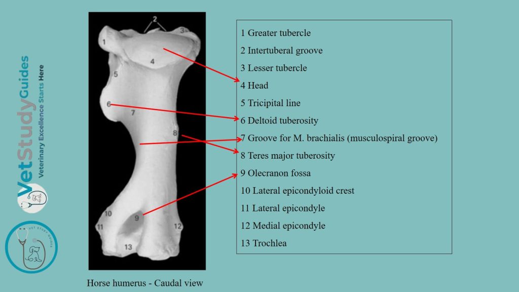
This structure bears above its middle the deltoid tuberosity/tuberositas deltoidea. From the latter, a rough line curves upward and backward to the lateral surface of the neck. It gives origin to the lateral head of the triceps muscle.
Below the tuberosity, the border inclines forward, becomes less salient, and ends at the coronoid fossa.
The posterior surface/facies caudalis is rounded from side to side and smooth.
Proximal extremity of the horse’s humerus
The proximal extremity/extremitas proximalis consists of the head, neck, two tuberosities, and the intertuberal groove.
Head of the humerus
The head/caput humeri presents an almost circular convex articular surface. It is about twice as extensive as the glenoid cavity of the scapula with which it articulates. In front of the head is a fossa, in which are several foramina.
Neck of the humerus
The neck/collum humeri is well defined behind, but is practically absent elsewhere.
Lateral and medial tuberosities of the humerus
Laterla tuberosity: The lateral tuberosity/tuberositas lateralis is placed antero-laterally. It consists of two defined parts: anterior and posterior.
The anterior part forms the lateral boundary of the intertuberal or bicipital groove. It gives attachment to the lateral branch of the supraspinatus muscle.
The posterior part gives attachment to the short insertion of the infraspinatus. However, its outer surface is coated with cartilage. The chief tendon of the infraspinatus muscle passes to be inserted into a triangular rough area below the anterior part.
Medial tuberosity: The medial tuberosity/tuberositas medialis is less salient. It also consists of anterior and posterior parts.
Here, the anterior part forms the medial boundary of the bicipital/intertuberal groove. It furnishes insertion to the inner branch of the supraspinatus above, and the posterior deep pectoral muscle below.
Again, the posterior part gives attachment to the subscapularis muscle.
Intertuberal/ bicipital groove
The intertuberal or bicipital groove/sulcus intertuberalis is situated in front. The anterior parts of the tuberosities bound it.
However, it is subdivided by an intermediate ridge. The groove is covered in the fresh state by cartilage and lodges the tendon of origin of the biceps brachii muscle.
Just below the intermediate ridge is a small fossa in which several foramina open.
Distal extremity of the horse’s humerus
The distal extremity has an oblique surface for articulation with the radius and ulna. This extremity consists of two condyles of very unequal size, separated by a ridge.
Medial condyle: The medial condyle/condylus medialis is much larger. It is crossed by a sagittal groove, on the anterior part of which there is usually a synovial fossa.
Posteriorly, the groove extends upward considerably above the rest of the articular surface. It reaches the olecranon fossa, and this part articulates with the trochlear/semilunar notch of the ulna.
Lateral and medial condyles
The lateral condyle/condylus lateralis is much smaller and is placed somewhat lower and further back. It gives the extremity an oblique appearance.
A wide, shallow groove marks it. The coronoid fossa/fossa coronoidea is situated in front, above the groove on the medial condyle.
It furnishes origin to part of the extensor carpi. Lateral to this fossa is a rough depression from which the common digital extensor arises.
Lateral and medial epicondyles
Behind and above the condyles are two thick ridges, the epicondyles.
Medial epicondyle: The medial epicondyle/epicondylus medialis is the more salient. It furnishes origin to the flexor muscles of the carpus and digit. Again, it also bears a tubercle for the attachment of the medial ligament of the elbow joint.
Lateral epicondyle: The lateral epicondyle/epicondylus lateralis bears laterally the condyloid crest/crista condyloidea. It forms here the outer boundary of the musculospiral groove, and gives origin to the extensor carpi radialis.
Below this is a rough excavation in which the lateral ligament is attached. The distal border of the epicondyle gives attachment to the ulnaris lateralis.
Between the epicondyles is the deep olecranon fossa/fossa olecrani, into which the processes anconseus projects.
Development of the horse humerus bone
The humerus ossifies from six centers: three primary centers for the shaft and extremities, and three secondary centers for the lateral tuberosity, the deltoid tuberosity, and the medial condyle, respectively.
The proximal end fuses with the shaft at about three and one-half years, the distal at about one and a half years of age.
FAQ’s on horse’s humerus
The proper anatomical neck/collum anatomicum is, however, indicated by the shallow depression which separates the head from the tuberosities, and gives attachment to the joint capsule.
The term tubercle, as used in human anatomy, does not apply well to the domestic animals.
The name intertuberal is designative of the position of the groove, while the term bicipital has reference to its occupation by the tendon of the biceps brachii. The term sulcus intertubercularis is also in common use.
Conclusion
Thus, the horse humerus is a long bone that consists of a cylindrical body and two extremities. Here, the body consists of 4 surfaces: lateral, medial, anterior, and posterior.
However, the proximal and distal extremities of the equine/horse’s humerus possess different identifying osteological features.
References
- Sisson, S., Anatomy of the domestic animals. W B Saunders Company, USA.
- Gabriele Barros Mothé, Anselmé Dutra, and Francisco Mendes Junior,
- Dyce and Wensing, Textbook of Veterinary Anatomy, 4th edition, Saunders, USA.
- de Alcântara Leite dos Reis, D., Gouveia, B.L.R., Júnior, J.C.R. et al. Comparative evaluation of anatomical details of thoracic limb bones of a horse to those of models produced via scanning and 3D printing. 3D Print Med 5, 13 (2019).
- Anna Dee Fails and Christianne Magee, Anatomy and physiology of the Farm Animals, 111 River Street, Hoboken, NJ 07030, USA.
- Hilary M. Clayton, Peter F. Flood, Diana S. Rosenstein, and David Mandeville, Clinical Anatomy of the Horse, First edition 2005, ISBN 07234 3302 X.
- Pasquini and Spurgeon, Anatomy of domestic animals, systemic and regional approaches.
- Victoria Aspinall B, and Melanie Cappello, Introduction to Veterinary Anatomy and Physiology Textbook, ISBN 978-0-7020-5735-9, Elsevier.