The horse metacarpal bones are the long type of bones in its skeleton. Here, you will learn the details of the large and small metacarpals from the forelimb of a horse.
Number and types of horse metacarpal bones
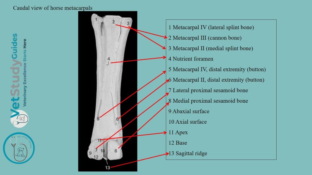
Three metacarpal bones are present in the horse. Of these, only one, the third or large metacarpal bone, is fully developed. This developed metacarpal carries a digit.
The other two, the second and fourth, are much reduced in the horse. They are commonly called the small metacarpal or splint bones.
The third/large metacarpal bone of the horse
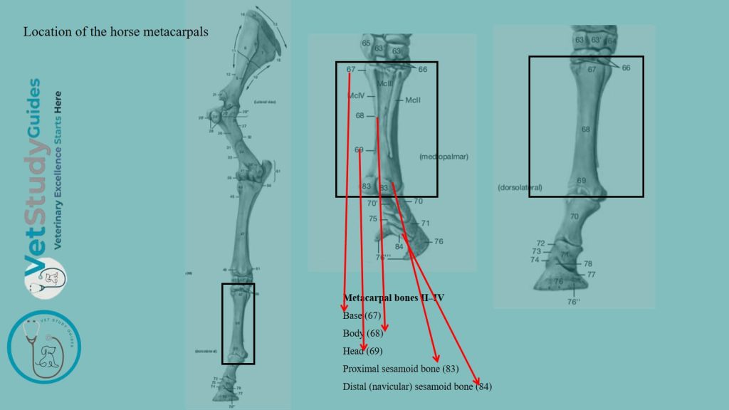
Location and type of bone: The third/ large metacarpal of a horse is a very strong, long bone. It is situated vertically between the carpus and the first phalanx. It consists of a shaft and two extremities: proximal and distal.
The shaft of the horse’s large metacarpal
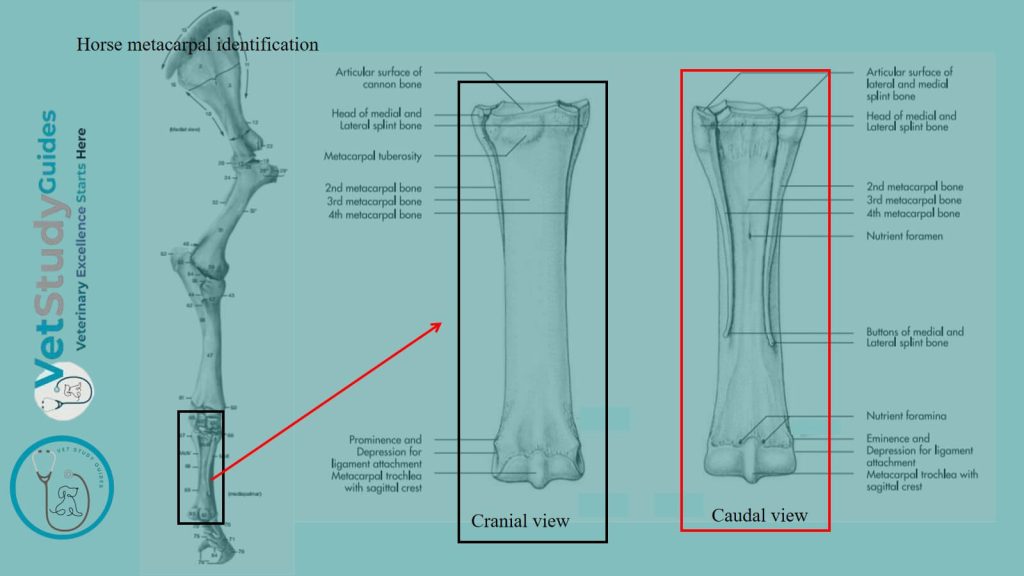
The shaft/corpus/body is semicylindrical and presents two surfaces and two borders. Here, the surfaces are dorsal and volar, and the borders are lateral and medial.
The dorsal surface is smooth, convex from side to side, and nearly straight in its length. The volar surface is somewhat convex from side to side, and with the small bones. It forms a wide groove which lodges the suspensory ligament.
On either side of its proximal two-thirds, it is roughened for the attachment of the small metacarpal bones. The nutrient foramen occurs at the junction of the proximal and middle thirds.
The distal part is wider and flattened. The borders are rounded.
Proximal extremity of the horse/equine metacarpals
The proximal extremity bears an undulating articular surface adapted to the distal row of carpal bones. The greater part supports the third carpal bone.
Again, the oblique lateral part is separated from the preceding by a ridge. It articulates with the fourth, and a small facet for the second is usually found at the medio-volar/caudomedial angle.
On either side is a notch separating two small facets. They articulate with the proximal ends of the small metacarpal bones.
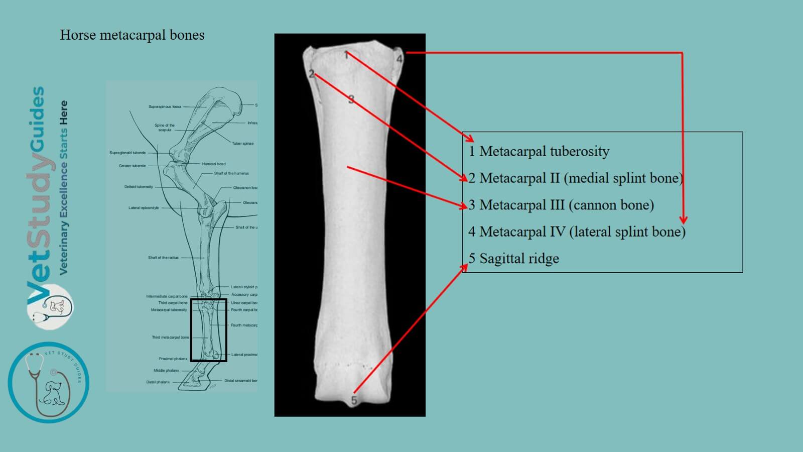
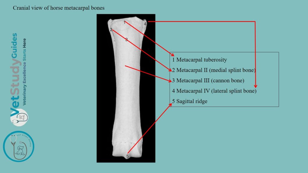
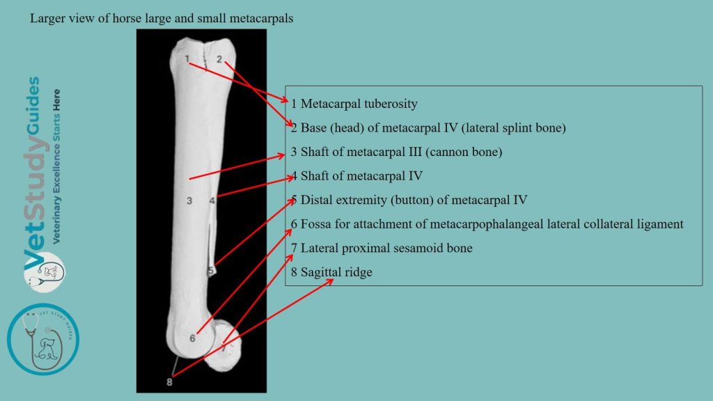
Toward the medial side of the dorsal surface is the metacarpal tuberosity. Within this structure, the extensor carpi radialis is inserted. However, the volar surface is roughened for the attachment of the suspensory ligament.
Distal extremity of the horse metacarpal bones
The distal extremity presents an articular surface for the first/proximal phalanx and the proximal sesamoid bones. This part of the metacarpal is composed of two condyles: lateral and medial.
A sagittal ridge separates these condyles. Here, the medial condyle is slightly larger in the horse metacarpal bone.
On either side is a small fossa, surmounted by a tubercle, for the attachment of the collateral ligaments of the fetlock joint.
Small metacarpal bones of the horse
These are, numerically, the second and fourth metacarpal bones in the horse. They are situated on either side of the volar surface of the large metacarpal bone.
They form the sides of the metacarpal groove. Each of the small metacarpals of a horse consists of a shaft and two extremities.
The shaft/body of the horse’s small metacarpal
The shaft/body/corpus is three-sided and tapers to the distal end. It is variably curved, convex toward the middle line of the limb.
The attached surface is flattened and is rough, except in its lower part. It is attached to the large metacarpal bone by an interosseous ligament, except near the distal end.
The dorsal/abaxial surface is smooth and rounded from side to side above, grooved below. The volar/axial surface is smooth and concave from edge to edge, except below. Below, it forms a rounded edge.
Extremities of the horse’s small metacarpal bones
Proximal extremity: The proximal extremity is relatively large in the horse’s small metacarpals. In the case of the medial bone, it usually bears two facets above which support the second and third carpal bones.
Again, the lateral small metacarpal bone has a single facet for articulation with the fourth carpal bone.
Each has also two facets for articulation with the large metacarpal. Again, it is elsewhere roughened for the attachment of ligaments and muscles. The medial bone may present a small facet behind the first carpal bone.
Distal extremity: The distal extremity is usually a small nodule, which projects to a variable extent in different subjects. It is easily felt in the living animal. This structure is situated two-thirds to three-fourths of the way down the region.
Development of the horse metacarpal bone
The large metacarpal bone of a horse ossifies from three centers. Here, the proximal extremity unites with the shaft before birth. However, the distal extremity is toward the middle of the second year.
The small metacarpal bones of a horse ossify from two centers, one of which is for the proximal extremity. Their distal ends are cartilaginous at birth.
Fusion of the middle part of the shaft with the large metacarpal bone is common.
FAQ on the horse’s large and small metacarpal bones
The large metacarpal is one of the strongest bones in the skeleton. The compact substance is especially thick in front and medially. The medullary cavity extends further toward the ends than in most of the long bones of the horse, and there is little spongy substance.
The small metacarpal bones vary greatly in length, thickness, and curvature. In the majority of cases, the medial bone is longer.
In other subjects, the lateral one is the longer, or there is no material difference. Sometimes the curvature is very pronounced, so that the distal end causes a decided projection.
The distal end is very variable in size and may be a mere point. In other cases, especially in large draft horses, it may present a prolongation which is regarded as the vestige of the digital skeleton.
Conclusion
So, the features of the large and small metacarpal bones of a horse present the features of a long bone. Its shaft has a semicylindrical appearance with unique dorsal and volar surfaces.
However, the extremities of the horse metacarpals present special osteological features for articulation with the carpus above and the phalanges below.
References
- Sisson, S., Anatomy of the domestic animals. W B Saunders Company, USA.
- Gabriele Barros Mothé, Anselmé Dutra, and Francisco Mendes Junior,
- Dyce and Wensing, Textbook of Veterinary Anatomy, 4th edition, Saunders, USA.
- Brokken, M. and Tucker, R. (2010). The metacarpal/metatarsal region. In Equine MRI, R.C. Murray (Ed.)
- de Alcântara Leite dos Reis, D., Gouveia, B.L.R., Júnior, J.C.R. et al. Comparative evaluation of anatomical details of thoracic limb bones of a horse to those of models produced via scanning and 3D printing. 3D Print Med 5, 13 (2019).
- Anna Dee Fails and Christianne Magee, Anatomy and physiology of the Farm Animals, 111 River Street, Hoboken, NJ 07030, USA.
- Eren, G., López-Albors, O., López Corbalán, M., & Latorre, R. (2026). Three-Dimensional Reconstruction of the Equine Palmar Metacarpal Region Using E12 Plastinated Sections. Animals: an open access journal from MDPI, 16(3), 449.
- Hilary M. Clayton, Peter F. Flood, Diana S. Rosenstein, and David Mandeville, Clinical Anatomy of the Horse, First edition 2005, ISBN 07234 3302 X.
- Pasquini and Spurgeon, Anatomy of domestic animals, systemic and regional approaches.
- Victoria Aspinall B, and Melanie Cappello, Introduction to Veterinary Anatomy and Physiology Textbook, ISBN 978-0-7020-5735-9, Elsevier.