The horse carpus contributes to forming the manus segment of the forelimb. They are the type of short bones of the horse skeleton.
In this guide, you will learn the details of the osteological features of the horse’s carpus.
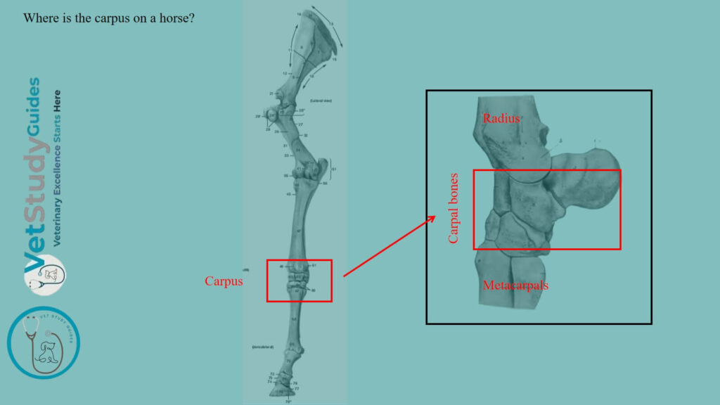
Where is the carpus on a horse?
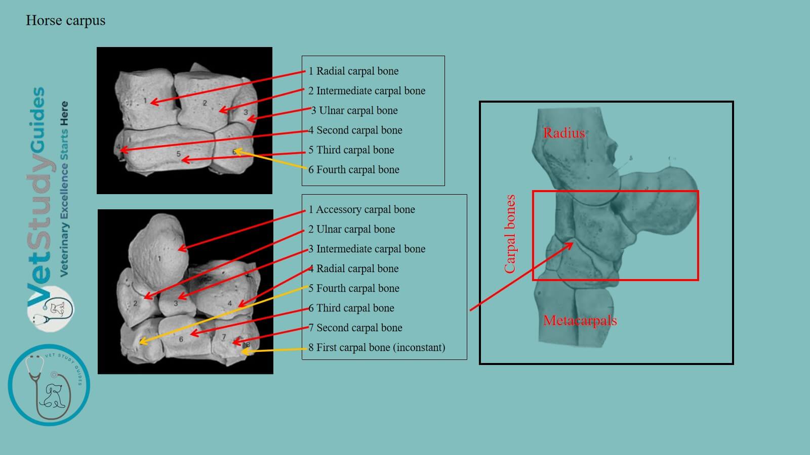
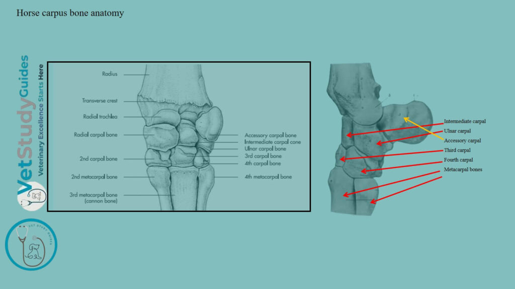
The horse’s carpus is located between the distal extremity of the radius and ulna, and the proximal extremity of the metacarpal bones. In the horse, it consists of seven or eight carpal bones/ossa carpi.
These carpal bones of the horses are arranged in two rows: proximal or antibrachial and distal or metacarpal.
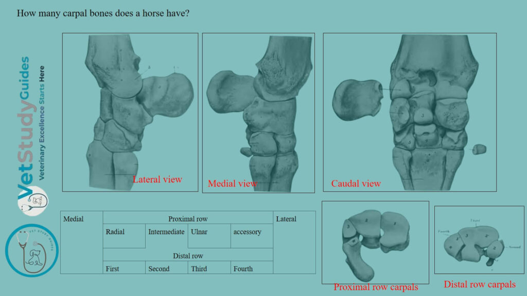
How many carpal bones does a horse have?
Thus, the horse has seven or eight (7/8) carpal bones. The names and relative positions of the carpal bones of the carpus are indicated below (Table 1)
| Medial | Proximal row | Lateral | |||
| Radial | Intermediate | Ulnar | accessory | ||
| Distal row | |||||
| First | Second | Third | Fourth | ||
Horse carpus
So, the term horse carpus means the total area that is formed by the seven or eight carpal bones. First, let’s see the overview of the horse’s carpus within a minute.
Overview: Here, the bones of the carpus, exclusive of the accessory, form an irregular quadrangular mass. The width of this carpal is about twice the height or the dorsovolar diameter.
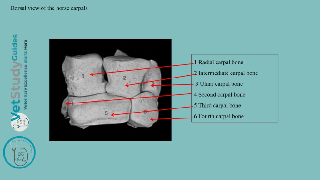
Dorsal surface: The dorsal surface is convex from side to side. It is depressed along the line of junction of the two rows and prominent below.
Volar surface: The volar surface is, in general, slightly convex but very irregular. It forms with the accessory carpal the carpal groove.
This surface is rendered smooth by the volar ligament. However, it is converted into the carpal canal/canalis carpi for the flexor tendons by the transverse carpal ligament. It stretches across from the accessory bone to the medial side.
Proximal surface: The proximal surface is widest medially and is elevated in front, concave behind. It is entirely articular and adapted to the carpal articular surface of the radius.
The distal surface is also articular and is irregularly faceted in adaptation to the surfaces of the metacarpal bones. Here, each of the distal bones usually articulates with two metacarpal bones. Sometimes the third rests on the third metacarpal only.
Medial and lateral surfaces: The medial and lateral surfaces are both irregular and rough, the former being wider. With the exception of the accessory, ulnar, and second, each bone articulates with two bones of the other row.
Horse carpal bones anatomy
Now, I will describe the individual carpal bones of the horse’s carpus with their unique features. Thus, you will easily identify these carpal bones from the horse’s carpus.
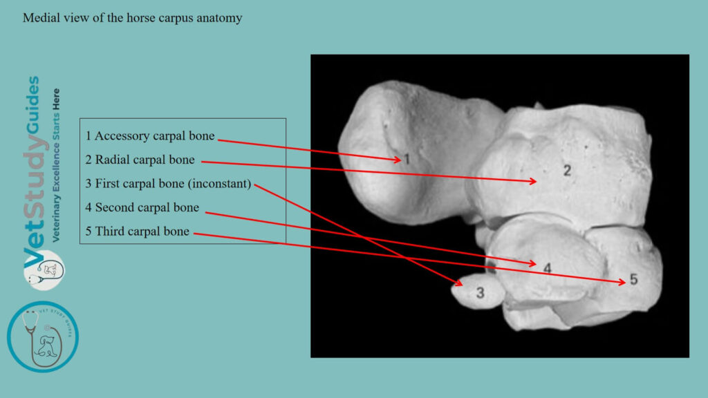
Radial carpal bone of a horse
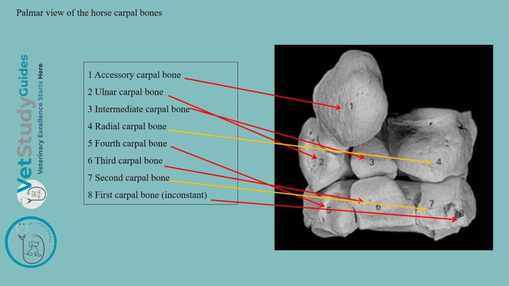
The radial carpal bone/os carpi radiale is the largest bone of the proximal row. It is somewhat compressed transversely and is clearly six-sided.
Proximal surface: The proximal surface is convex in front, concave behind. It articulates with the medial facet on the distal end of the radius.
Distal surface: The distal surface is also convex in front and concave behind. It articulates with the second and third carpal bones.
Lateral surface: The lateral surface bears upper and lower facets on its anterior part for articulation with the intermediate. Between and behind the lateral surface, it is excavated and rough.
Dorsal surface: The dorsal surface is rough and slightly convex. The medial surface and the volar surface are rough and tuberculate.

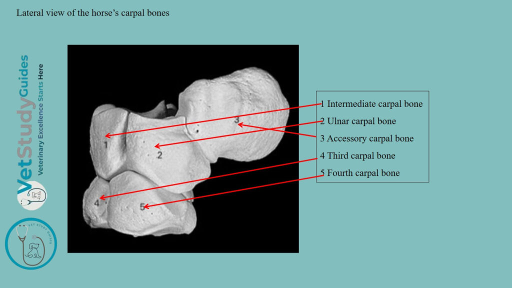
Intermediate carpal bone of the horse
The horse’s intermediate carpal bone/os carpi intermedium is somewhat wedge-shaped, wider in front than behind.
Proximal surface: The proximal surface of the intermediate carpal bone is saddle-shaped. It articulates with the middle facet on the distal end of the radius.
Distal surface: The distal surface is smaller, convex in front, and concave behind. It articulates with the third and fourth carpal bones.
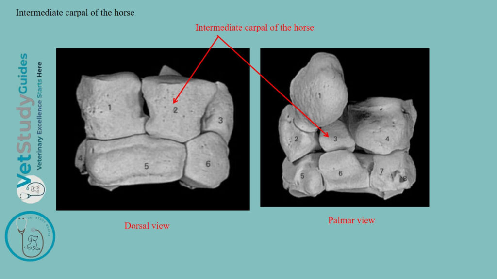
Medial surface: The medial surface has upper and lower facets for articulation with the radial carpal. Between these facets, it is excavated and rough.
Lateral surface: The lateral surface is similar to the medial surface. However, this surface articulates with the horse’s ulnar carpal.
Dorsal surface: The dorsal surface is rough and slightly convex.
Volar surface: The volar surface bears a tuberosity on its lower part.
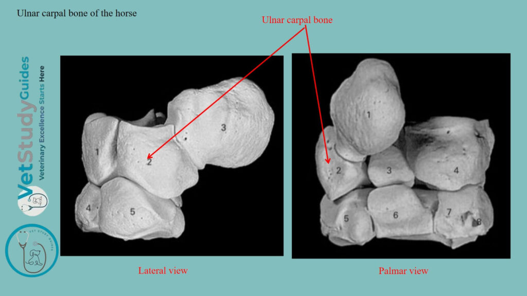
Horse ulnar carpal bone anatomy
The Horse’s ulnar carpal bone/os carpi ulnare is the smallest and most irregular bone of the proximal row.
Proximal surface: The proximal surface is concave and fits the lower part of the lateral facet on the distal end of the radius.
Distal surface: The distal surface is oblique and undulating for articulation with the fourth carpal bone.
Medial surface: The medial surface has upper and lower facets for articulation with the intermediate.
Dorsal and lateral surfaces: The dorsal and lateral surfaces are continuous, convex, and rough.
Volar surface: The volar surface is oblique and bears a concave facet (small articular surface) for articulation with the accessory carpal bone.
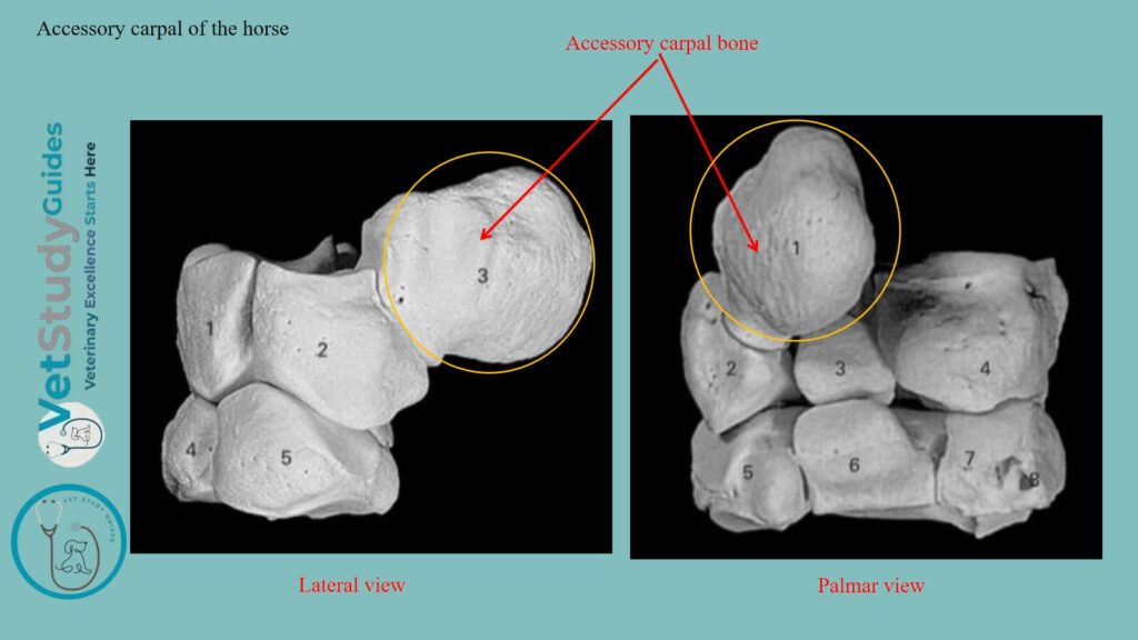
Accessory carpal bone of a horse
The horse’s accessory carpal bone/os carpi accessorium is situated behind the ulnar carpal bone and the lateral part of the distal end of the radius. It is discoid and presents for description two surfaces and a circumference.
Medial surface: The medial surface is concave and forms the lateral wall of the carpal groove.
Lateral surface: The lateral surface is convex and rough. A smooth groove for a tendon crosses its anterior part obliquely downward and slightly forward.
Dorsal border: The dorsal border bears two facets. Here, the proximal one is concave and articulates with the back of the lateral facet on the distal end of the radius. However, the distal one is convex and articulates with the ulnar carpal bone. The remainder of the circumference is rounded and rough.
Exceptions: The accessory does not directly bear weight and may be regarded as a sesamoid bone. It is interposed in the course of the tendons of the middle and lateral flexors of the carpus.
Posterior border: The posterior border furnishes attachment to the transverse carpal ligament. It completes the carpal canal for the flexors of the digit.
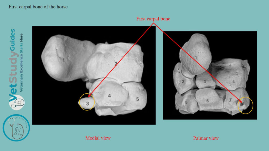
Horse first carpal from the carpus
The first carpal bone/os carpale primum, is a small, inconstant bone. It is commonly about the size and shape of a pea.
Location: This first carpal is situated in the distal part of the medial ligament of the carpus, behind the second carpal bone. This bone appears to be absent on both sides in about half of the cases.
However, in a good many subjects, it is present on one side only. In size, it varies from a minute nodule to a discoid or cylindrical mass 12-15 mm in length.
Exceptions: In exceptional cases it articulates with both the second carpal and the second metacarpal bone. In other cases with the former only, but in the majority of bones no articular facet is present.
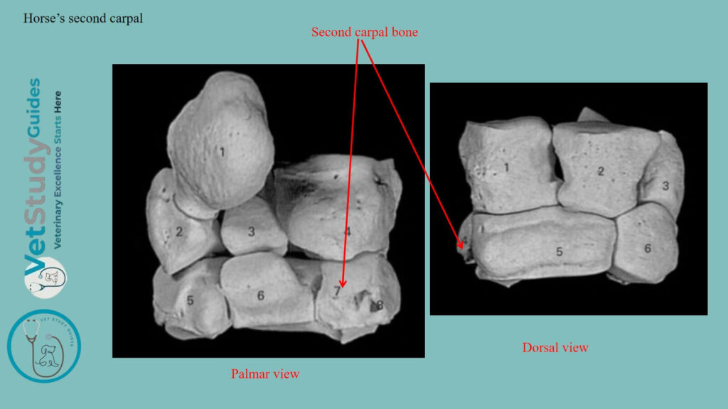
Horse’s second carpal bone
The horse’s second carpal bone/os carpale secundum is the smallest constant bone of the distal row. It is irregularly hemispherical in shape.
Proximal surface: The proximal surface is a convex facet that is continued upon the volar surface and articulates with the radial carpal.
Lateral surface: The lateral surface faces obliquely outward and forward. It bears three facets for articulation with the third carpal bone.
Dorsal and medial surfaces: The dorsal and medial surfaces are continuous and bear a tuberosity to which the collateral ligament is attached.
Distal surface: The distal surface is articular and consists of a large flattened facet for the second or inner metacarpal bone. It also has a small one for the third or large metacarpal bone.
Volar surface: Some bones have a small facet on the lower part of the volar surface, which articulates with the first carpal bone.
The third carpal bone of horse anatomy
The horse’s third carpal bone/os carpale tertium is much the largest bone of the distal row. It forms more than two-thirds of the width of the latter.
This third carpal is flattened from above downward, and is twice as wide in front as behind.
Proximal surface: The proximal surface consists of two facets separated by an anteroposterior ridge. Again, the medial facet is concave and articulates with the radial carpal.
However, the lateral facet for the intermediate carpal is concave in front and convex behind.
Distal surface: The distal surface is slightly undulating and articulates almost entirely with the third or large metacarpal bone.
But it usually bears a small oblique facet at its medial side for the second metacarpal. However, there is commonly a non-articular depression laterally.
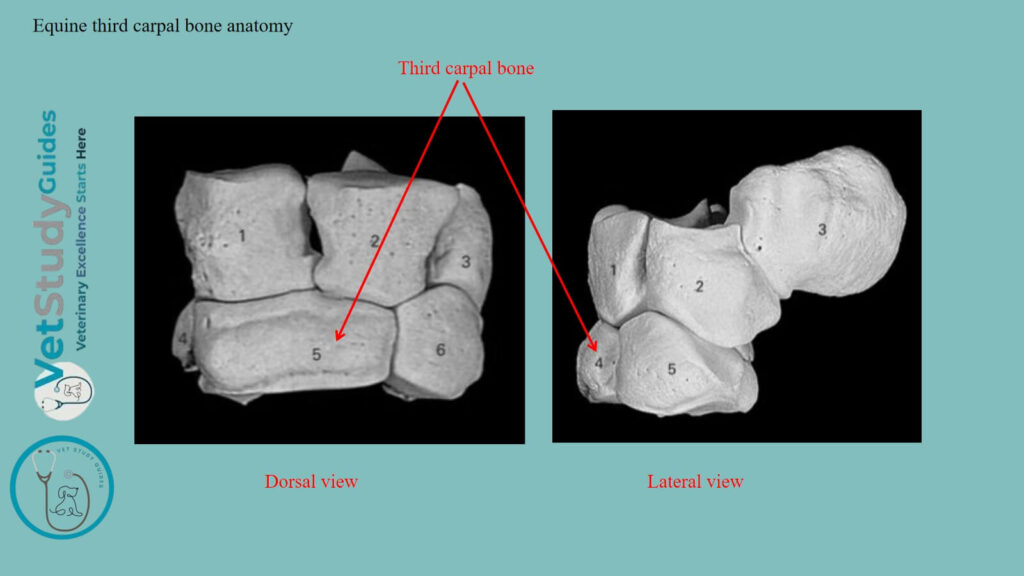
Medial surface: The medial surface faces backward and inward. It bears three facets for articulation with the second carpal, between which it is excavated and rough.
Lateral surface: The lateral surface has two facets for articulation with the fourth carpal. It is depressed and rough in its middle.
Dorsal surface: The dorsal surface is convex and is crossed by a rough transverse ridge.
Volar surface: The volar surface is relatively small and is rounded. Its upper part is encroached upon by the proximal articular surface, below which it is rough.
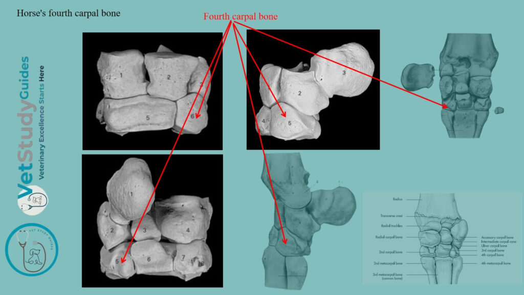
Horse’s fourth carpal
The horse’s fourth carpal bone/os carpale quartum is somewhat wedge-shaped. It is readily distinguished from the second by its greater size and its volar tubercle.
Proximal surface: The proximal surface articulates with the intermediate and ulnar. It is convex and curves outward, backward, and downward, encroaching on the lateral and volar surfaces.
Distal surface: The distal surface bears two medial facets for the third or large metacarpal and a lateral one for the fourth or lateral metacarpal bone.
Medial surface: The medial surface has two or three facets for articulation with the third carpal, between which it is excavated and rough.
Dorsal surface: The dorsal surface is convex and rough.
Lateral surface: The lateral surface is small, being encroached upon by the proximal articular surface.
Volar surface: The volar bears a tubercle on its distal part.
FAQ’s in horse carpus
The bones of the carpus, exclusive of the accessory, form an irregular quadrangular mass. The width of the horse’s carpus is about twice the height or the dorso-volar diameter.
The dorsal surface is convex from side to side, depressed along the line of junction of the two rows, and prominent below. The volar surface is, in general, slightly convex but very irregular.
Conclusion
So, the horse carpus is formed by the proximal and distal rows of carpal bones. Here, the proximal row of the horse’s carpus consists of 4 bones. Whereas, the distal row of the carpus presents 3/4 bones.
References
- Sisson, S., Anatomy of the domestic animals. W B Saunders Company, USA.
- Dyce and Wensing, Textbook of Veterinary Anatomy, 4th edition, Saunders, USA.
- Anna Dee Fails and Christianne Magee, Anatomy and physiology of the Farm Animals, 111 River Street, Hoboken, NJ 07030, USA.
- Oheida, A. H., Shalgum, A. A., Alrtib, A. M., Booker, A. O., Ben-Naser, K. M. & Davies,. H. M. (2023). Variation in palmaromedial articulations of carpometacarpal joints in Thoroughbred and Standardbred racehorses. Open Veterinary Journal, 13 (5), 569-575.
- Solounias, N., Danowitz, M., Stachtiaris, E., Khurana, A., Araim, M., Sayegh, M., & Natale, J. (2018). Evolutionary anatomy of the horse’s manus. Royal Society Open Science, 5(1), 171782.
- Hilary M. Clayton, Peter F. Flood, Diana S. Rosenstein, and David Mandeville, Clinical Anatomy of the Horse, First edition 2005, ISBN 07234 3302 X.
- Gabriele Barros Mothé, Anselmé Dutra, and Francisco Mendes Junior, Descriptive osteology of the equine thoracic limb. RCMOS – Multidisciplinary Scientific Journal of O Saber. ISSN: 2675-9128. São Paulo-SP, Year IV, v.1, n.1, Jan./Jul. 2024.
- Pasquini and Spurgeon, Anatomy of domestic animals, systemic and regional approaches.
- Hagenbach, et all., (2024). Anatomical structures in the horse carpal. Frontiers in Veterinary Science, 11, 1431777. https://doi.org/10.3389/fvets.2024.1431777
- Victoria Aspinall B, and Melanie Cappello, Introduction to Veterinary Anatomy and Physiology Textbook, ISBN 978-0-7020-5735-9, Elsevier.
- Simon, V. and Dyson, S.J. (2010), Radiological anatomic variation of the carpus in horses with carpal lameness and control horses. Veterinary Radiology & Ultrasound, 51: 601-606.