
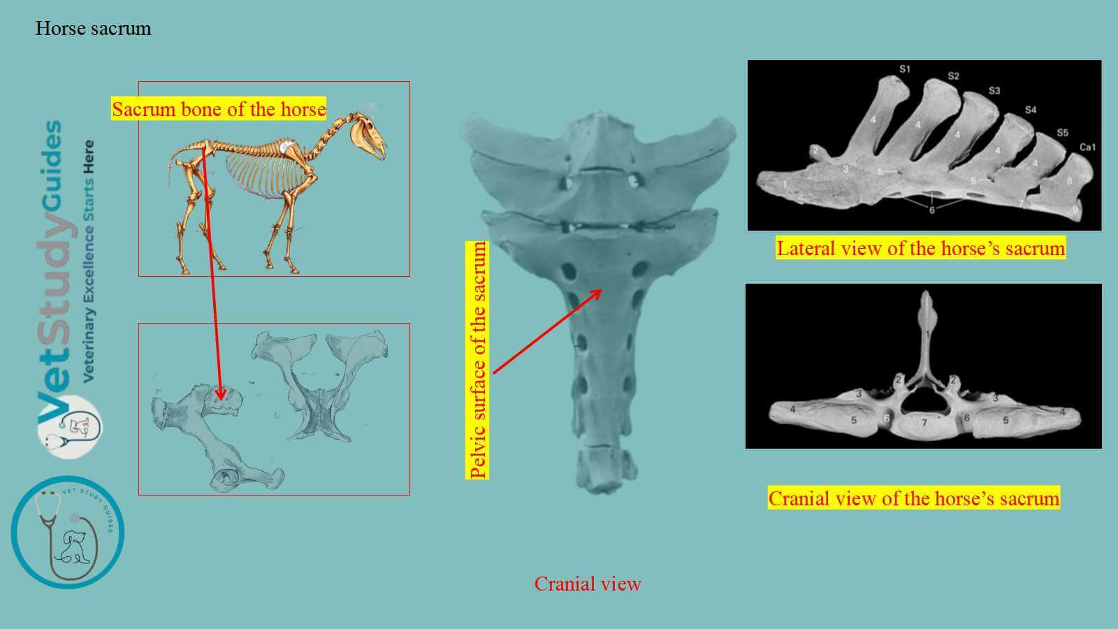
The horse sacrum forms from the fusion of five sacral vertebrae. It is conveniently described as a single bone of the vertebral column.
Here, I will describe the osteological features of the horse’s sacrum with diagrams. However, this sacrum, or os sacrum, is also known as the false vertebra in the axial skeleton.
Where is the sacrum on a horse?
The figure shows the location of the horse’s sacrum on its skeleton. It is located along the midline of the skeleton (vertebral column) between the lumbar and coccygeal/caudal vertebrae.
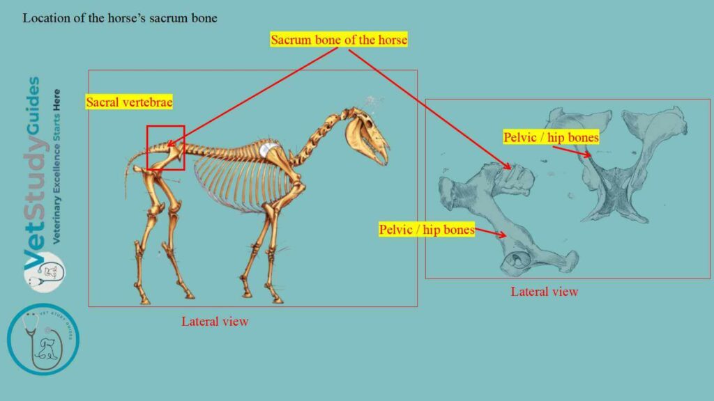
It also shows that five sacral vertebrae fuse to form a single sacrum in the equine. However, this bone has exceptional osteological features compared with the sacrum of the ox, sheep, goat, and dog.
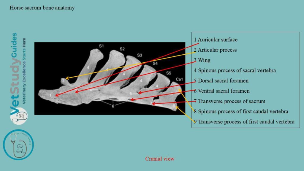
Horse sacrum bone anatomy
The horse’s sacrum is triangular and wedged between the ilia. Here, the ilia of the sacrum articulate very firmly on each side.
The long axis of the sacrum is gently curved and slightly oblique. Thus, the posterior end is a little higher than the anterior end.
It presents two surfaces, two borders, a base, and an apex.
Surfaces and borders of the horse’s sacrum
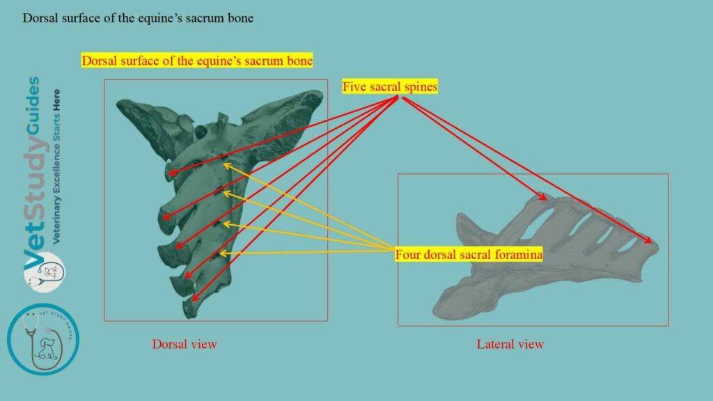
Dorsal surface: The dorsal surface presents the five sacral spines centrally. They point upward and backward and have (except the first) tuberous summits that are sometimes bifid.
On either side of the spines is a groove that contains the four dorsal sacral foramina. Here, the dorsal branches of the sacral nerves emerge through these foramina.
Pelvic surface: The pelvic surface is concave in its length, wide in front, narrow behind. The curvature is variable and is more pronounced in the mare than in the stallion.
However, it is marked by four more or less distinct transverse lines. They indicate the demarcation of the bodies of the vertebrae.
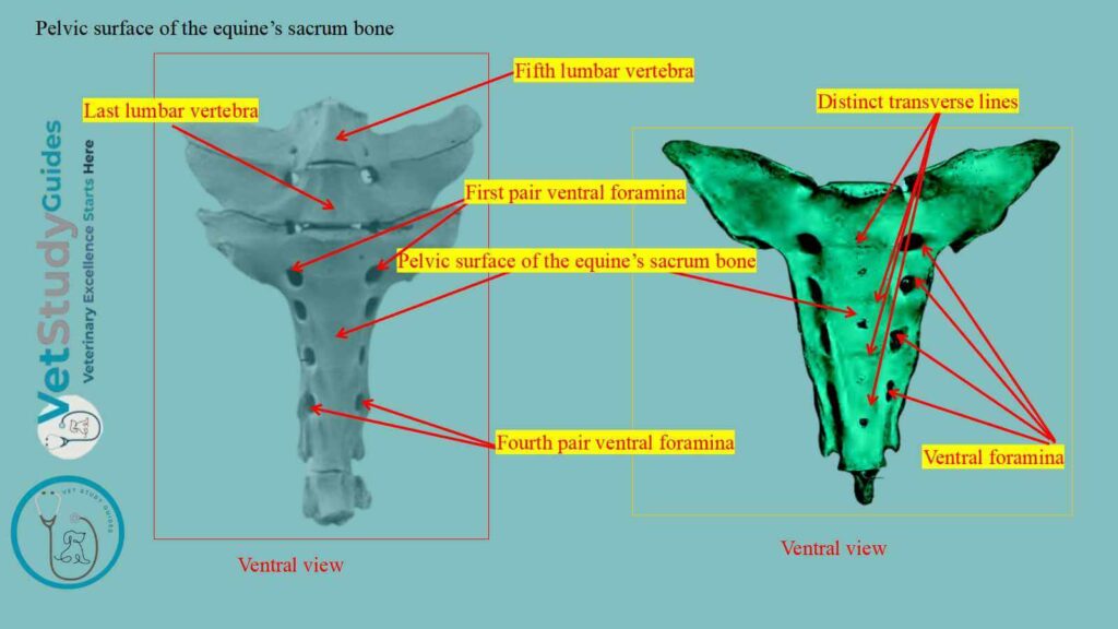
At the ends of these lines are the ventral sacral foramina. However, they are larger than the dorsal series and diminish in size from first to last.
They transmit the ventral divisions of the horse’s sacral nerves. However, the dorsal and ventral foramina communicate with the sacral canal.
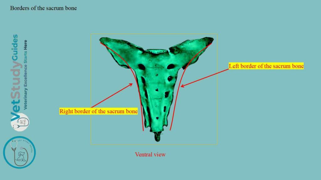
Lateral borders: The lateral borders (right and left) of the equine’s sacrum are rough, thick in front, and thin behind.
Base of the equine’s sacrum bone
The horse sacrum has a base that is directed forward and is relatively wide. It presents the body of the first sacral segment centrally (first sacral vertebra).
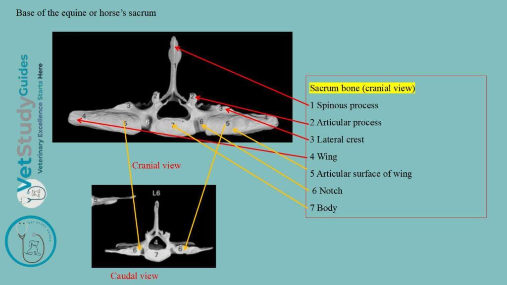
- Body of the base: Here, the body is wide transversely, flattened dorso-ventrally, and has a rounded surface. Again, this surface articulates with the last lumbar vertebra through an intervertebral fibrocartilage.
- Margin of the body: The ventral margin projects slightly, forming the promontory.
- Notch and foramen: On either side of the body of the equine sacrum, there is a smooth notch. This notch, with one on the last lumbar vertebra, forms a large foramen. Within this foramen, the ventral branch of the last lumbar nerve passes.
- Articular processes: Above the body is the entrance to the sacral canal, flanked by a pair of articular processes. These processes project upward and forward from the arch, and have concave surfaces medially for articulation with those of the last lumbar vertebra.
Lateral to each is a smooth notch that becomes a foramen by apposition with the last lumbar vertebra.
Exceptional oval articular surfaces: The lateral parts of the base, the alae or wings, are strong prismatic masses with pointed ends. Each has in front a large, oval, slightly convex surface for articulation with the transverse process of the last lumbar vertebra.
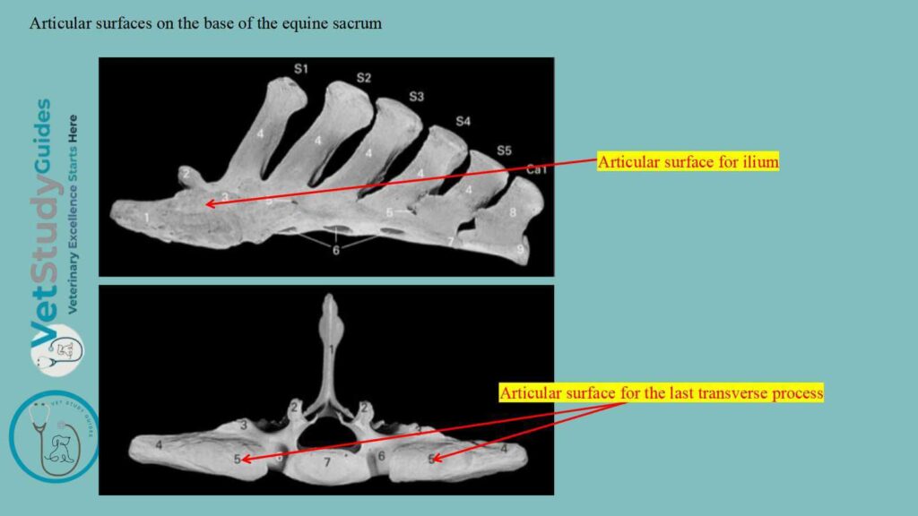
Articular surface for ilium: Posteriorly there is an elongated oval area which faces dorso-laterally. This is the auricular surface, which articulates with the ilium. However, it is slightly concave in its length, and somewhat rough and irregular.
The rest of the dorsal surface of the wing is roughened for ligamentous attachment, while the ventral surface is smooth.
Apex of the equine/horse’s sacrum
The apex is the posterior aspect of the last sacral vertebra and is quite small. It presents an elliptical, flattened surface of the body of the vertebra.
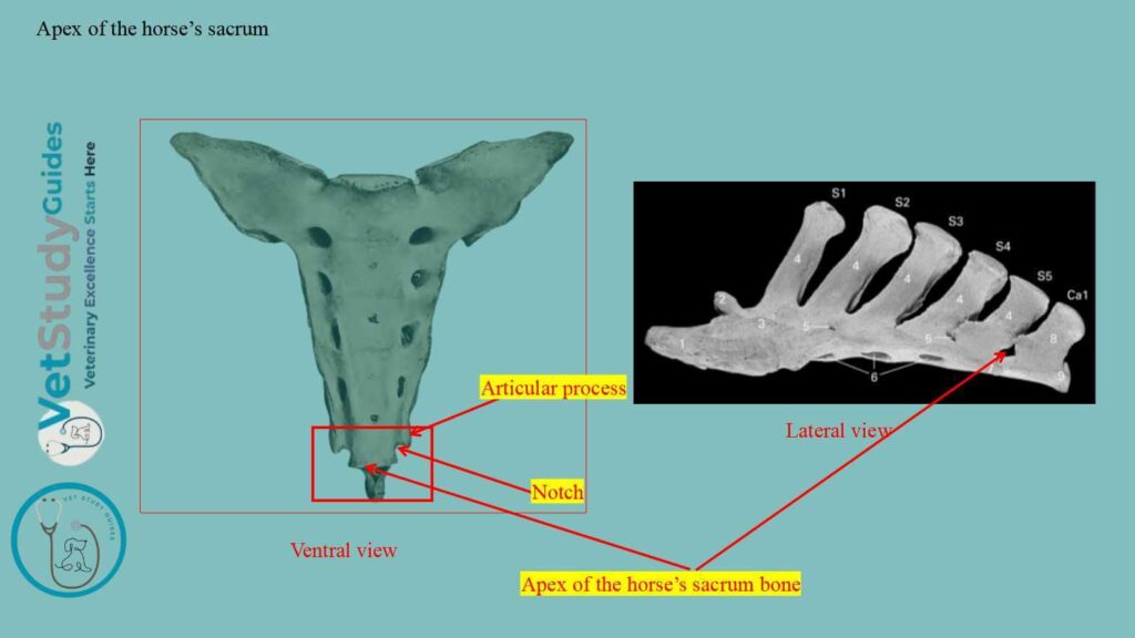
Triangular opening: Just above the body, there is the triangular posterior opening of the sacral canal. However, the last sacral spine surrounds this triangular opening.
Notches and articular processes: A pair of narrow notches exists between the arch and body of the sacrum (apex). Just above this portion, rudiments of articular processes may occur.
What is the horse’s sacral canal?
The term horse’s sacral canal refers to the part of the vertebral canal that traverses the sacrum.
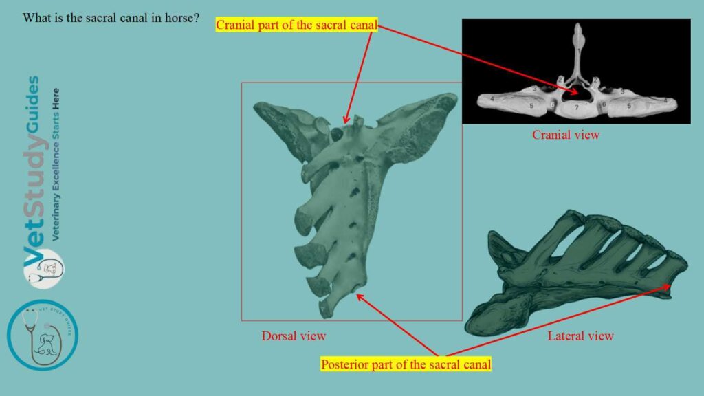
- Anterior part: It is large and triangular, with rounded angles.
- Height and width: However, its width is nearly twice its height.
- Posterior part: Traced backward, it diminishes rapidly in size, and the posterior opening is small and triangular.
The term lateral part designates the portion lateral to the foramina, which results from the fusion of the transverse processes.
FAQ’s on the horse’s sacrum bone anatomy
The several sacral vertebrae ossify in the typical manner. Separate centers for costal elements in the lateral parts have not yet been found in domesticated animals.
Here, fusion begins in front and is usually not complete until older age. The lateral parts unite before the bodies. It is rather curious that the epiphyseal plates of adjacent segments unite before they fuse with the main portion of the bodies.
The first spine is relatively thin and narrow, and is not so high as the sacral angle of the ilium. The second is the longest and highest, and the length and height diminish to the last. The bases of the spines are often fused in old subjects.
Conclusion
Thus, the horse sacrum has distinct spinous processes due to incomplete fusion. However, the ventral surface of the sacrum is less concave than in ruminants or other species.
Again, the lateral sacral crests of the equine (horse) sacrum are ill-developed. The wings are pointed and prismatic and contain an exceptional oval articular surface for the last lumbar vertebra.
References
- Sisson, S., Anatomy of the domestic animals. W. B. Saunders Company, USA.
- Dyce and Wensing, Textbook of Veterinary Anatomy, 4th edition, Saunders, USA.
- Goff, L. M., et al., (2006). Movement between the equine ilium and sacrum, Equine Veterinary Journal. Supplement, (36), 457–461.
- Beth Vanhorn and Robert W. Clark, Veterinary Assisting: Fundamentals & Applications, ISBN-13: 978-1-4354-5387-6, Maxwell Drive, Clifton Park, NY 12065-2919, USA.
- Anna Dee Fails and Christianne Magee, Anatomy and physiology of the Farm Animals, 111 River Street, Hoboken, NJ 07030, USA.
- Dalin, G. and Jeffcott, L.B. (1986), Sacroiliac Joint of the Horse 1. Gross morphology. Anatomia, Histologia, Embryologia, 15: 80-94.
- Pasquini and Spurgeon, Anatomy of domestic animals, systemic and regional approaches.
- Hilary M. Clayton, Peter F. Flood, Diana S. Rosenstein, and David Mandeville, Clinical Anatomy of the Horse, First edition 2005, ISBN 07234 3302 X.
- Ogden NKE, et al., 2025. Computed tomography of the equine caudal spine and pelvis. Equine Vet J; Sep;57(5):1279-1289.
- Victoria Aspinall B and Melanie Cappello, Introduction to Veterinary Anatomy and Physiology Textbook, ISBN 978-0-7020-5735-9, Elsevier.