
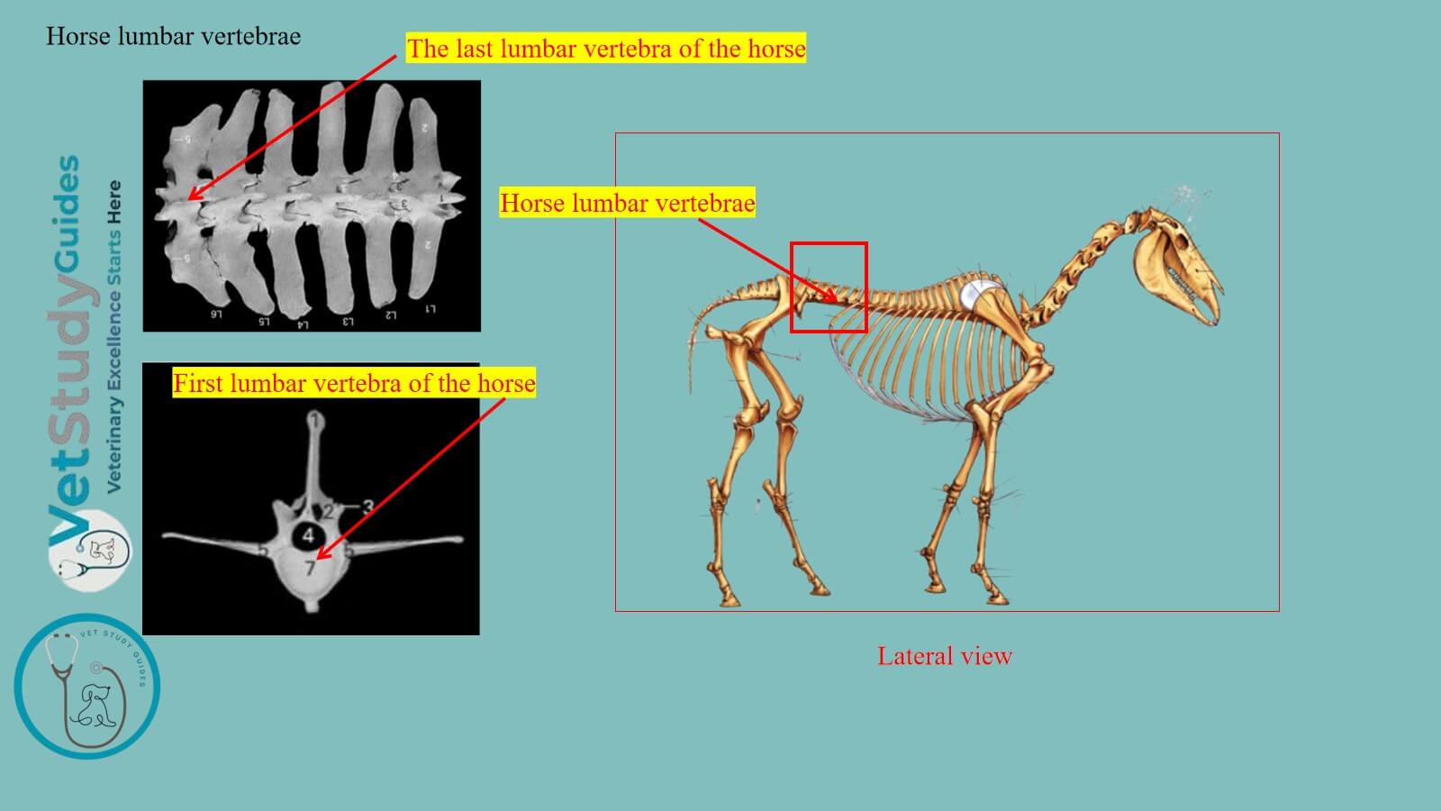
Horse lumbar vertebrae with their bodies and long transverse processes from the bony roof of the abdomen. They also contribute to forming the horse’s vertebral column along the axial skeleton.
Here, I will discuss the osteological features of the horse’s lumbar vertebrae with the diagrams. However, you will know the exceptional features of the last lumbar vertebra of the horse compared to those of other animals.
Where are the horse’s lumbar vertebrae?
The figure shows the location of the horse’s lumbar vertebrae from its axial skeleton. They are located between the horse’s thoracic vertebrae and sacrum bone.
However, the wider bodies and the longer transverse processes of the horse’s lumbar vertebrae form the roof of the abdomen. Different abdominal muscles of the horse arise or insert on the transverse processes of these vertebrae.

How many lumbar vertebrae does a horse have?
The figure shows and identifies the total number of the horse’s lumbar vertebrae from the skeleton.
Number: The horse’s lumbar vertebrae are usually six in number (Lumbar 1 – Lumbar 6). They are characterized by the loner size and form of their transverse processes.
However, the number of the horse’s lumbar vertebrae is expressed with ‘L’. Thus, these vertebrae are: L1, L2, L3, L4, L5, and L6.

Horse lumbar vertebrae anatomy
To describe the anatomy of the equine/horse’s lumbar vertebrae, you might know the typical features. A typical vertebra or lumbar vertebra of a horse or equine has the following structures –
- Wide and long body: It possesses extremities and surfaces (like the typical one)
- Arches: There are also right and left lateral arches; each arch consists of the ventral pedicle and dorsal lamina (shown in the diagram), and
- Processes: They possess paired cranial and caudal articular processes, a single spinous process, and longer lateral paired transverse processes.
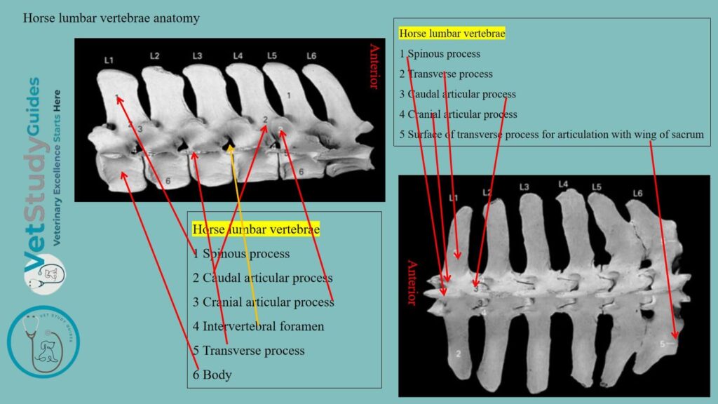
However, before going into a detailed description of the equine lumbar vertebrae, I would like to summarize their features in a table –
| Horse’s lumbar vertebrae | Features |
| Bodies | Wider and longer in the horse |
| Arches (pedicle and lamina) | Pedicles are comparatively short and notchedThey form the larger intervertebral foramina |
| Transverse processes | Long and in the form of a bony plate They inclines ventrally |
| Spinous processes | They are in the form of a quadrilateral plate |
| Articular processes | Both cranial and caudal articular processes are well-developed |
Okay, now let’s see all the features of the equine lumbar vertebra with diagrams.
Bodies of the horse’s lumbar vertebrae
The bodies of the horse’s lumbar vertebrae are comparatively wider and longer than those of others. Here, the bodies of these vertebrae present the anterior and posterior extremities.
The convexity and concavity of the anterior and posterior extremities are not prominent, respectively. However, these features are more prominent in the horse’s cervical vertebrae anatomy.
Here, the bodies of the first three are semi-elliptical on cross-section. However, they also present a distinct ventral crest.

From the fourth backward, the horse’s lumbar becomes wider and flatter. However, the ventral crest of these lumbar vertebrae (L4-L6) subsides.
Arches of the equine lumbar vertebrae
Here, the equine lumbar vertebrae exhibit right- and left-sided arches. Each right and left arch of these vertebrae shows the pedicle and lamina.

Size of arches: The arches of the first three are about equal in size . However, they are similar to those of the equine’s last thoracic vertebrae.
Behind the third vertebra, the remaining lumbar vertebrae increase in breadth and height.
Notches on the arches: Both arches present the cranial and caudal notches. However, the posterior notches are much deeper than the anterior ones.
Processes of the horse’s lumbar
Like the typical vertebra, the horse’s lumbar also presents three types of processes –
- Anterior and posterior paired articular processes,
- Longer transverse processes, and
- Single upward-directed single spinous process,
Articular processes of the equine lumbar
Anterior articular process of the horse’s lumbar: The anterior articular processes are fused with the mammillary processes in the horse. However, they present dorsally concave surfaces for articulation with the posterior pair of the preceding vertebra.

Posterior articular processes: The posterior articular processes project distinctly from the arch at the base of the spinous process. However, they have ventrally convex articular surfaces. It fit into the concave surfaces of the anterior pair of vertebrae.
Transverse processes of the horse’s lumbar
The transverse processes are elongated plates, flattened dorso-ventrally in the horse.
Direction: They project outward and usually curve slightly downward.
Length: The length of the equine lumbar’s transverse process increases to the third or fourth. Then it diminishes to the last, which is the shortest.
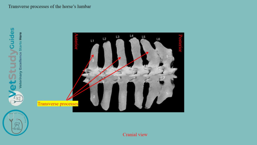
Serial changes of the transverse process in the horse lumbar
First one or two vertebrae: The first one or two usually curve somewhat backward, the last two decidedly forward.
Fifth lumbar: Those of the fifth have an oval concave facet on the medial side of the posterior border. However, it is designed for articulation with the sixth process.
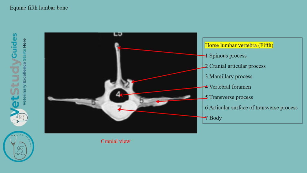
Sixth lumbar (Exceptional): The sixth lumbar has a corresponding convex facet on the anterior border. However, it also has a larger concave surface on the posterior border for articulation with the wing of the sacrum.
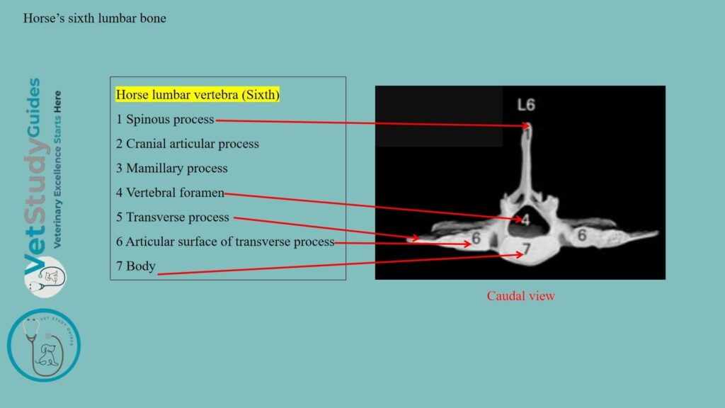
Exceptional features for the fifth lumbar: Sometimes the fifth process has a small surface for articulation with the fourth. The medial part of the sixth process is thick, the lateral part thinner, narrower, and curved forward.
The medial part of the fifth is also somewhat thickened. Medial to the articular surfaces, the edges of the transverse processes are cut into by notches. They form foramina by apposition with each other and the sacrum.
Spinous process of the horse’s lumbar vertebrae
The spinous processes resemble those of the last two thoracic vertebrae. However, they are usually about equal in height, but minor differences are common. Again, the width diminishes in the last three lumbar vertebrae.

Neural ring and foramina
The neural rings in the equine are rounded up to the third lumbar vertebrae. However, they gradually become triangular in shape after the third lumbar vertebra.
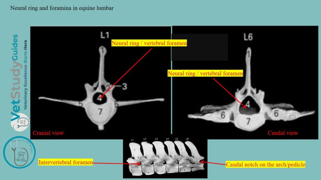
The intervertebral foramina in the adjacent lumbar vertebrae are larger. Sometimes, the anterior few foramina of some of the equine’s lumbar vertebrae are double.
FAQ’s on the horse’s lumbar vertebrae
The occurrence of a rib in connection with the transverse process of the first lumbar vertebra is common. A reduction to five has been observed frequently.
However, it may or may not be compensated for by an additional thoracic vertebra. Very few horses have seven lumbar vertebrae, especially with the normal thoracic number. An anomalous vertebra with mixed thoracic and lumbar characters sometimes occurs at the junction of the two regions.
The transverse processes of the ox lumbar vertebrae incline cranially. Whereas it is inclined ventrally in the equine lumbar vertebrae. However, the summits of the spinous processes of the ox lumbar vertebrae are sometimes bifid.
Conclusion
So, the horse lumbar vertebrae show exceptional osteological features in their bodies and transverse processes. Here, the bodies of the horse’s lumbar vertebrae are wider and compressed dorso-ventrally.
However, the longer lateral paired transverse processes are unique for the horse’s lumbar vertebrae. Again, the upward-directed quadrilateral spinous process is also a unique feature in this lumbar vertebra.
References
- Sisson, S., Anatomy of the domestic animals. W B Saunders Company, USA.
- Dyce and Wensing, Textbook of Veterinary Anatomy, 4th edition, Saunders, USA.
- Spoormakers TJP, et al., Pathologic changes in the lumbar region of the equine vertebral column, Equi. Vet J. 2024; 56(5): 1031–1040.
- Beth Vanhorn and Robert W. Clark, Veterinary Assisting: Fundamentals & Applications, ISBN-13: 978-1-4354-5387-6, Maxwell Drive, Clifton Park, NY 12065-2919 USA.
- Anna Dee Fails and Christianne Magee, Anatomy and physiology of the Farm Animals, 111 River Street, Hoboken, NJ 07030, USA.
- Hilary M. Clayton, Peter F. Flood, Diana S. Rosenstein, and David Mandeville, Clinical Anatomy of the Horse, First edition 2005, ISBN 07234 3302 X.
- Zaneb et al. (2013) “Functional anatomy and biomechanics of the equine thoracolumbar spine: a review,” Tur. Jr of Vet. and Ani Sci., Vol. 37: No. 4, Article 2. https://doi.org/10.3906/vet-1205-45.
- Pasquini and Spurgeon, Anatomy of domestic animals, systemic and regional approaches.
- Victoria Aspinall B, and Melanie Cappello, Introduction to Veterinary Anatomy and Physiology Textbook, ISBN 978-0-7020-5735-9, Elsevier.
- Scilimati, N., et al. (2023). Computed Tomographic (CT) Features of the Most Caudal Lumbar Vertebrae. Animals, 13(4), 743. link: https://doi.org/10.3390/ani13040743
Reviewed and Edited by