
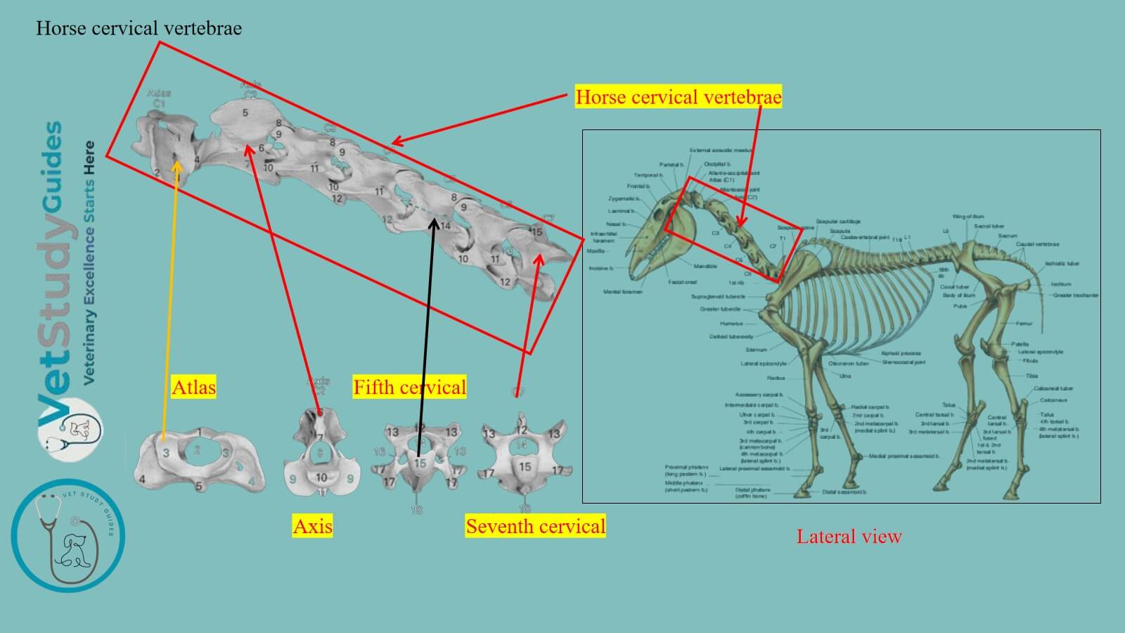
The cervical vertebrae of a horse form the neck skeleton. Here, in this guide, I will describe the osteological features of the horse’s cervical vertebrae with diagrams.
The cervical vertebrae contribute to the formation of the axial skeleton. However, they are characterized by the presence of short processes of various shapes and a foramen transversarium.
Horse cervical vertebrae
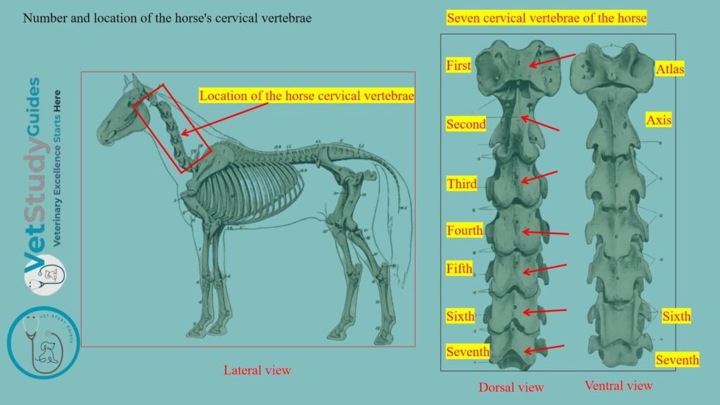
Number: The cervical vertebrae are seven in number in a horse. Here, the figure shows the location, number, and features of the horse’s cervical vertebrae.
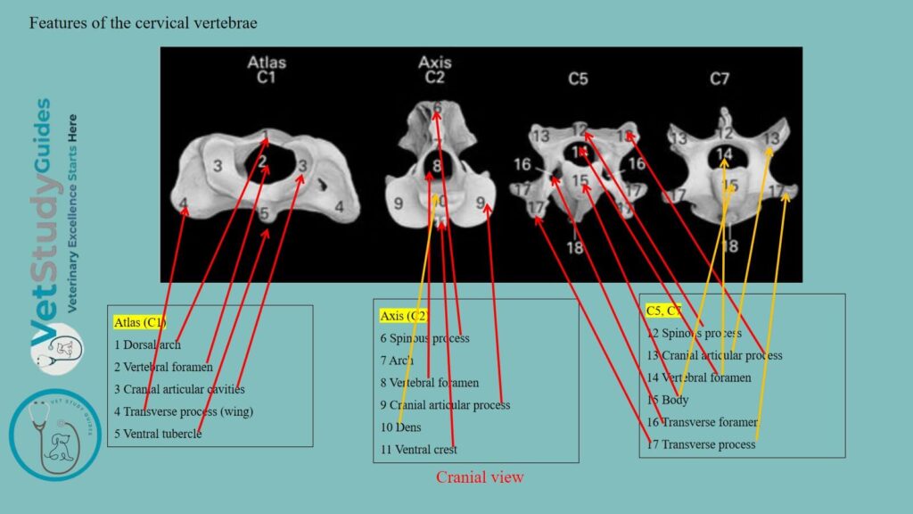
Features of the cervical vertebrae
First and second cervical: The first and second cervical vertebrae are highly modified in conformity with the special functions of support and movements of the head.
Sixth and seventh cervical bones: The sixth and seventh present special characters, but do not differ greatly from the typical.
With the exception of the first, they are quadrangular, massive, and longer than the vertebrae of other regions. However, they decrease in length from the second to the last.
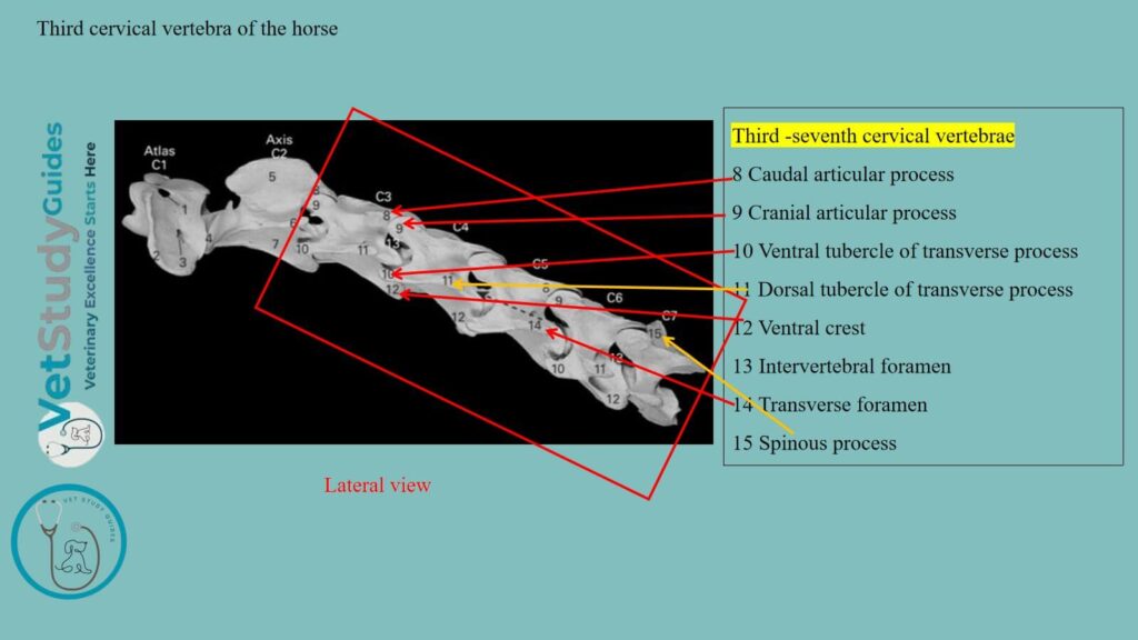
Third, fourth, and fifth cervical vertebrae: The third, fourth, and fifth have the features of a typical vertebra.
Structure of a typical cervical vertebra in a horse
A typical vertebra (for example, a horse’s third, fourth, and fifth cervical vertebrae) possesses a body, arches, and processes. Here, the body is cylindrical and contains dorsal and ventral surfaces.
Again, the lateral and medial arches (identified in the figure) possess a ventral pedicle and dorsal lamina. However, there are three processes (spinous, transverse, and articular) in the horse’s cervical vertebrae.
Here, the articular processes again divide into cranial and caudal paired divisions. You might learn the details of these bodies, arches, and processes from the horse’s typical vertebra (cervical vertebra).
I am going to describe all these features of the horse’s typical cervical vertebrae in detail. However, you may also use this same template of description for other vertebrae, like the thoracic and lumbar vertebrae.

The body of the cervical vertebrae (third, fourth, and fifth) in the horse
The body is longer than that of other vertebrae. It presents two surfaces: ventral and dorsal surfaces, and two extremities: anterior and posterior.
Ventral surface: Here, the ventral surface presents a median ventral spine. It becomes more prominent as it is traced backward. However, it is tuberculate at its posterior end and separates two concave areas.
Dorsal surface: The dorsal surface has a flat central area, which is narrow in the middle of the vertebrae. It widens at either end and attaches to the dorsal longitudinal ligament.
Grooves: On either side of the dorsal area, there is a groove that lodges the longitudinal spinal vein. These lateral grooves are connected about the middle of the surface by a transverse furrow. There are several foramina through which veins emerge from the spongy substance of the body.
Anterior extremity: The anterior extremity, or head, has an oval articular surface that faces forward and downward. It is strongly convex and wider above than below.
Posterior extremity: The posterior extremity is larger and has a nearly circular cotyloid cavity.
Arches of the horse cervical vertebrae anatomy
The arches are on the right and left, and each is large and strong. It is perforated on either side by a foramen which communicates with the foramen transversarium.
Notches: The vertebral notches on the arches are large. Notches of the corresponding cervical vertebrae form the intervertebral foramina in the horse’s vertebral column.
Processes of the cervical or typical vertebra in the horse
I will describe the three types of processes of the horse’s typical vertebrae separately. However, the features of these three processes vary in shape and size across different vertebrae of the vertebral column.
Articular processes
The articular processes are large. Their articular surfaces are extensive, oval in outline, and slightly concave. Here, the anterior ones are directed dorso-medially; the posterior, ventro-laterally.
The remaining surface is mainly roughened for ligamentous and muscular attachment. A crest connects the articular processes of the same side on the fourth and fifth vertebrae. However, on the third, it does not reach the anterior process.
Transverse processes
The transverse processes are large and plate-like in the horse’s cervical vertebra. Each arises by two roots, one from the arch and one from the body.
Foramen transversariium: Between the roots, there is the foramen transversariium, through which the vertebral vessels and a nerve pass. The aggregate of these foramina constitutes the canalis transversarius.
The process divides laterally into anterior and posterior branches. These branches are thickened and rough for muscular attachment.
Spinous process
The spinous process is a low, narrow crest that arises from the junction of the laminae. They widen behind and are connected by ridges with the posterior articular processes.
Thus, these are the typical features of the horse’s cervical vertebrae (third, fourth, and fifth). Now, you may easily learn the features of the other cervical or even thoracic, lumbar, sacral, and caudal vertebrae by comparing them with these typical vertebrae.
Horse atlas bone
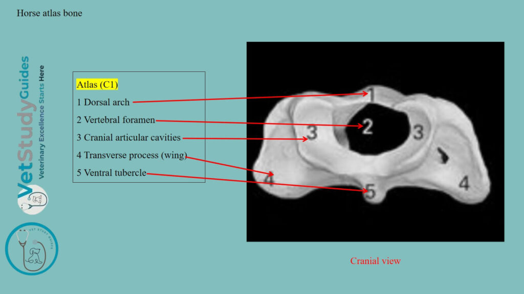
The first cervical vertebra/atlas of a horse is decidedly atypical in form and structure.
Exceptions: The body and spinous process are absent. It has the form of a strong ring, from which two curved plates, the wings, project laterally.
Rings of the horse atlas
The ring encloses a very large vertebral foramen and consists of two lateral masses connected by dorsal and ventral arches.
Lateral masses: The lateral masses present two deep oval anterior articular cavities, which receive the occipital condyles. They are separated by a wide notch above and a narrow one below.
The lateral margin is notched, and a triangular non-articular depression cuts into the medial part of each cavity.
The posterior articular surfaces are somewhat saddle-shaped. They are confluent at the ventral arch but widely separated dorsally. However, they do not conform in shape to the corresponding surfaces of the axis.
Dorsal arch: The dorsal arch presents a median dorsal tubercle and is concave ventrally. It is perforated on either side near its anterior margin by the intervertebral foramen. However, the anterior border is deeply notched, and the posterior is thin and concave.
Ventral arch: The ventral arch is thicker, narrower, and less curved than the dorsal. On its lower surface is the ventral tubercle, into which the terminal tendon of the longus colli muscle is inserted.
Fovea dentis: The upper face has, posteriorly, a transversely concave articular surface, the fovea dentis. On this structure, the dens or odontoid process of the axis rests.
In front of this is a transverse rough excavation and a ridge for the attachment of the ligamentum dentis.
Wings/alae of the horse’s atlas bone
The wings or alae of the horse’s atlas are modified transverse processes. They are extensive curved plates which project ventro-laterally and backward from the lateral masses. Here, the dorsal surface is concave.
Fossa atlantis: Between the ventral aspect of the wing and the lateral mass is a cavity, the fossa atlantis. In this structure, there is a foramen that opens into the vertebral canal.
The border is thick and rough, and its position can be recognized in the living animal. Two foramina perforate each wing –
First: The anterior one, the foramen alare, is connected with the intervertebral foramen by a short groove.
Second: The posterior one is the foramen transversarium.
Horse axis bone
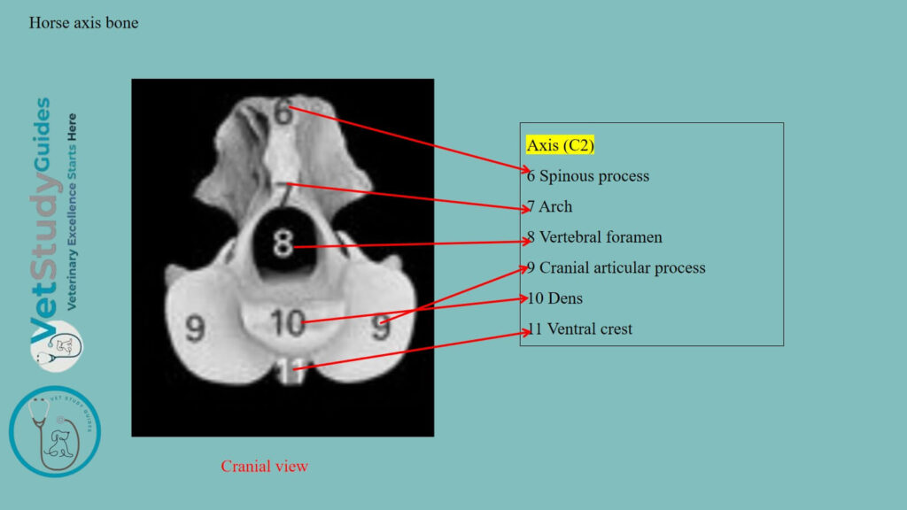
The axis is the longest of the vertebrae and is characterized by the presence of the dens or odontoid process. It projects from the anterior part of the body.
Extremities of the horse’s axis vertebra
Anterior extremity and odontoid process: The anterior extremity of the body presents centrally with the dens (odontoid process) or dens axis. It has a convex articular surface ventrally for articulation with the ventral arch of the atlas. It also possesses two rough depressions for the attachment of the ligamentum dentis dorsally.
Flanking this on either side are the modified anterior articular processes. They have saddle-shaped articular surfaces confluent ventrally with those of the dens.
Posterior extremity: The posterior extremity has the usual cavity. The ventral spine resembles that of the typical vertebrae.
Arches and processes of the horse axis
Arch: The arch presents a notch on each side of its anterior border in the young subject. It is formed into a foramen by a ligament that later ossifies. The posterior border has the usual notches.
Processes: The posterior articular processes are typical. The transverse processes are small, single, and project backward. The foramen transversarium is small.
The spinous process is very large and strong. Its free border is rough, thickens posteriorly. However, it continues to the articular processes by two ridges. The lateral surfaces are concave and rough for muscular attachment.
Sixth cervical vertebra in a horse
The sixth cervical vertebra has the following distinctive features –
Body: It is shorter and wider than the fifth.
Arch: The arch is large, especially posteriorly.

Processes: Posterior articular process: The posterior articular processes are shorter, thicker, and further apart. Each is connected with the corresponding anterior one by a thick ridge.
Spinous process: The spinous process is less rudimentary; it is half an inch or more in height.
Transverse process: The transverse processes have three branches; the third part is thick. However, it is almost a sagittal plate, which, with its fellow, forms a wide ventral groove with the body.
Again, the other branches correspond to those of the typical vertebrae, but are short and thicker.
Foramen transversarium: The foramen transversarium is large; below its posterior end, there is a fossa. The ventral spine is small and is less prominent posteriorly.
The seventh cervical vertebra in a horse
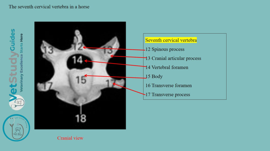
The seventh cervical vertebra is readily distinguished by the following characters –
Body: It is shorter and wider than the others. The body is flattened dorso-ventrally and wide, especially behind. Here, it has a facet on each side for articulation with the head of the first rib.
Arch: The arch and its notches are large.
Processes: The anterior articular processes are wider and longer than the posterior pair. The spinous process is an inch or more in height.
The transverse process is undivided and has no foramen transversarium. The ventral crest of the horse’s 7th cervical is replaced by a pair of tubercles.
FAQ’s in horse cervical vertebrae
The atlas ossifies from four centers: two for the ventral arch, and one on either side for each lateral mass, wing, and half of the dorsal arch.
At birth, the bone consists of three pieces: the ventral arch and two lateral parts, which are separated by a layer of cartilage in the dorsal median line and by two ventrolateral layers. These parts are usually fused at about six months.
The third branch of the transverse process and the fossa of some of the horse’s atlas are sometimes absent or reduced on one side. However, in some atlases of the horse, a large foramen transversarium is present on one side or (rarely) on both sides.
The axis has six or seven centers of ossification. In addition to the usual five, one or two appear for the dens, which are regarded as the displaced body of the atlas. A nucleus behind the dens, which remains distinct until three or four years of age, is considered to be the head of the axis.
Conclusion
So, the horse’s cervical vertebrae are seven in number and form the basis of the neck. Here, the third, fourth, and fifth possess the typical features of the horse’s vertebrae.
However, the first and second cervical vertebrae of the horses are highly modified for their adaptation. Again, the sixth and seventh cervical vertebrae also possess unique features (in body and process) compared to those of the typical vertebrae.
References
- Sisson, S., Anatomy of the domestic animals. W B Saunders Company, USA.
- Beth Vanhorn and Robert W. Clark, Veterinary Assisting: Fundamentals & Applications, ISBN-13: 978-1-4354-5387-6, Maxwell Drive, Clifton Park, NY 12065-2919 USA.
- Dyce and Wensing, Textbook of Veterinary Anatomy, 4th edition, Saunders, USA.
- Anna Dee Fails and Christianne Magee, Anatomy and physiology of the Farm Animals, 111 River Street, Hoboken, NJ 07030, USA.
- May-Davis, S., et al., (2023). Morphology of the Sixth Cervical Vertebra in the horse. Animals,13(10), 1672. https://doi.org/10.3390/ani13101672
- Hilary M. Clayton, Peter F. Flood, Diana S. Rosenstein, and David Mandeville, Clinical Anatomy of the Horse, First edition 2005, ISBN 07234 3302 X.
- Pasquini and Spurgeon, Anatomy of domestic animals, systemic and regional approaches.
- Victoria Aspinall B, and Melanie Cappello, Introduction to Veterinary Anatomy and Physiology Textbook, ISBN 978-0-7020-5735-9, Elsevier.
- Strootmann, T., et al., (2026). Anatomical Variations of the Ventral Lamina of the Sixth Cervical Vertebra in a horse. Animals, 16(3), 424.
Reviewed and Edited by