

The horse ribs or costae are elongated and curved modified bones. They form the skeleton of the lateral thoracic walls. However, they are arranged serially in pairs, each corresponding in number to a thoracic vertebra.
Here, I will describe the details of the horse’s rib anatomy with diagrams. However, you will know the types of ribs and the structure of a typical rib from the horse’s axial skeleton.
Where are a horse’s ribs?
Ribs are the part of the horse’s axial skeleton that forms the lateral thoracic wall of the thorax. Each rib articulates dorsally with two vertebrae and is continued ventrally to the sternum by a costal cartilage.
Here, the figure shows the location of the ribs and their contribution to forming the horse’s thorax.

Types of ribs in a horse
There are three different types of ribs in animals. However, they vary among horses, oxen, dogs, and other animals.
- Sternal ribs: Those ribs that articulate with the sternum by means of their cartilages are termed sternal ribs/costae sternales.
- Asternal ribs: The remaining ribs, which articulate with the sternum indirectly with other cartilages, are asternal ribs/costae asternales.
Floating ribs: Ribs at the end of the series that have their ventral ends free in the abdominal wall are named floating ribs/costae fiuctuantes.
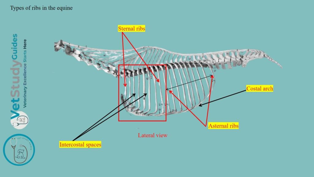
What is the intercostal space? Well, the intervals between the ribs are called intercostal spaces (spatia intercostalia). The figure shows the different types of ribs and intercostal spaces from the horse’s skeleton.
How many ribs are in a horse?
Number of ribs: There are usually eighteen pairs of ribs in the horse’s thorax. However, a nineteenth rib on one side or both is not at all rare.
Here, eight are sternal ribs, the remainder are asternal; however, there are no floating ribs in the horse’s skeleton.
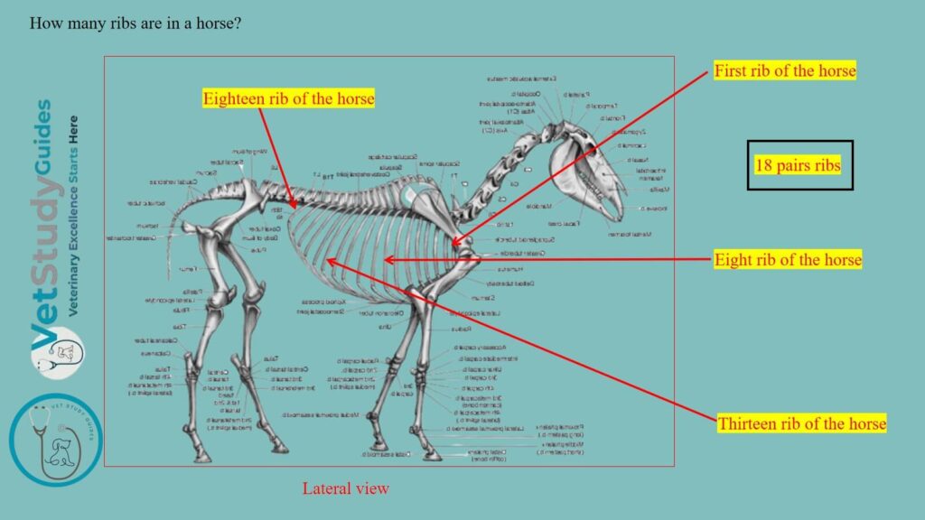
Ribs from different parts of the series vary greatly in length, curvature, and other characteristics. I will therefore consider as a type a rib from the middle of the series first, and afterward note the chief serial differences.
Typical rib of a horse
A typical rib of a horse consists of a shaft and two extremities. Here, the shaft of the rib has the following –
- Lateral and medial surfaces
- Anterior and posterior borders,
However, the rib shows the two extremities: the vertebral/proximal extremity and the sternal/distal extremity.

The shaft of the horse’s rib
The shaft of a horse rib is band-like and varies much in length, breadth, and curvature in different ribs. They are strongly curved, but the curvature is most pronounced in the dorsal third.
The ventral part of the horse’s rib is twisted and inclined inward. Thus, when a rib is laid with its lateral surface on the table, the sternal end is raised.
Lateral surface of the rib
The lateral surface is convex in both length and transverse direction. However, its anterior part is grooved longitudinally.
A distinct angle, that is, a point at which the curve of the rib changes rather suddenly. There is a rough elevation on the lateral surface that gives attachment to the longissimus muscle.

Medial surface of the rib
The medial surface is smooth and concave in its length. However, it is rounded on both sides.
Here, the costal groove/sulcus costse is situated posteriorly. However, it is very distinct above and fades out about the middle. Again, this groove of the rib contains the intercostal vein.
Borders of the ribs
The anterior border of a horse rib is concave, whereas the posterior border is convex. Here, the figure shows the borders of the horse’s ribs.
Extremities of the ribs
Vertebral extremity of the horse rib
The vertebral extremity consists of the head, neck, and tubercle. Here, the figure shows the vertebral and sternal extremities from the horse’s rib.
Head: The head/capitulum costse has an articular surface known as the facies articularis capituli costse. It is composed of two convex facets, anterior and posterior.
They are separated by a groove/sulcus capituli for the attachment of the conjugal ligament. It articulates with the cavity formed by facets on the bodies of two adjacent horse’s thoracic vertebrae and the intervertebral fibro-cartilage.
Neck: The neck/collar is roughened above and in front.
Tubercle: The tubercle/tuberculum costae is placed above and behind the junction of the neck and shaft. However, it has an oval surface, which is known as the facies articularis tuberculi costae. It is designed for articulation with the transverse process of the corresponding thoracic vertebra.

Sternal extremity of the horse’s rib
The sternal extremity/extremitas sternalis is slightly enlarged and is united with the costal cartilage.
Horse ribs anatomy
All eighteen ribs from the horse’s axial skeleton follow the typical features of the rib, with exceptions. However, the exceptional features of the different ribs are described below.

First rib of the horse
Appearances: The first rib is easily distinguished in the horse. It is the shortest, and the shaft widens greatly toward the sternal end. At the lower part of the anterior border, there is a smooth.
Tubercle: There is an impression where the brachial vein curves around it. Above this, there is commonly a small tubercle that indicates the lower limit of the insertion of the scalenus muscle.
Costal groove: The costal groove is absent in the horse’s first rib.
Head: The head is large and has two facets of unequal extent, which meet at an acute angle in front. Here, the smaller one faces forward and articulates with the last cervical vertebra. Again, the larger one is directed medially and articulates with the first thoracic vertebra.
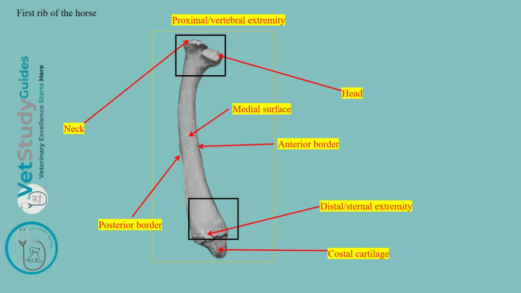
Neck: The neck is thick and very short. The tubercle is larger than that of any other rib and has an extensive articular surface which is convex in its length.
Sternal end: The sternal end is larger than that of any other rib. However, it is thick and very wide, and is slightly turned forward.
Last/ eighteen rib of the horse
The last rib is the most slender and regularly curved. It is usually, but a little longer than the second. The facet on the tubercle is confluent with that of the head.
This feature, however, is common on the seventeenth, and may occur on the sixteenth.
Serial position of the other ribs in equine
The serial position of the other ribs may be determined approximately by the following considerations –
Length: The length increases from the first to the tenth and eleventh, and then diminishes.
Width: The width increases somewhat to the sixth and then diminishes.
Anterior border: The anterior border is thin and sharp from the second to the eighth, and behind this, it becomes thick and rounded.
Groove: The groove of the lateral surface is distinct on the fourth to the eighth inclusive.
Curvature: The curvature increases rapidly from the second to the seventh. However, it continues the same until the sixteenth, and then decreases very noticeably.

Inclination: In regard to the dorso-ventral direction, the first rib inclines a little forward, the second is about vertical. However, behind this, they slope backward in increasing degree. Thus, a transverse plane tangent to the ventral ends of the last pair cuts the third lumbar vertebra.
Head: The head and tubercle diminish in size from first to last. Their relative positions change, in that the tubercle of the first rib is almost directly lateral to the head. Again, while further back it gradually comes to lie behind it.
Neck: The neck is longest on the longest ribs, and is absent on the last two or three. A costo-transverse foramen is formed between the neck and the transverse process.
Costal cartilages in the horse ribs
The first costal cartilage is an inch or more (2.5 to 3 cm.) in length. The dorsal end is very wide and thick, whereas the sternal end is small. The two articulate with each other as well as with the sternum.

Sternal and asternal rib cartilages: The cartilages of the other sternal ribs increase progressively in length and become more rounded.
The sternal end is enlarged and has an elliptical convex facet for articulation with the sternum. The cartilages of the asternal ribs are long, slender, and pointed.
The ninth is very firmly attached to the eighth. It and the next two are the longest, and behind this, they diminish progressively in size.
Costal arch: The cartilages of the asternal ribs of the horse overlap and are attached to each other by elastic tissue, forming the costal arch.

Exceptions: Except in the case of the first, the cartilage does not continue the direction of the rib. But forms with the latter an angle open in front, and increasing from second to last.
More or less extensive ossification is to be regarded as a normal occurrence, especially in the cartilages of the sternal ribs.
FAQ’s on horse rib anatomy
Well, A nineteenth rib on one side or both is not at all rare. It is usually imperfectly developed and quite variable. In many cases, it is a mere strip of cartilage connected by ligament with the first lumbar transverse process. However, in other cases, it is well developed and may be fused with the process.
It is commonly floating, but may be attached to the eighth costal cartilage.
Reduction in number is uncommon. In rare cases, the first rib is imperfectly developed and does not reach the sternum. Partial fusion of adjacent ribs and other anomalies occur.
The ribs ossify in cartilage from three centers, one each for the shaft, head, and tubercle. Here, the third center is absent in some of the posterior ribs.
Conclusion
So, the ribs of the horse have a body and two extremities. Here, the body of the horse’s rib consists of 2 surfaces, 2 borders, and a costal groove. However, the extremities of the horse’s ribs are proximal/vertebral and distal/sternal.
Here, the vertebral extremity of the horse’s rib possesses the head, neck, and tubercle. Again, the sternal extremity of the horse’s rib attaches to the sternum with costal cartilages.
References
- Sisson, S., Anatomy of the domestic animals. W B Saunders Company, USA.
- Dyce and Wensing, Textbook of Veterinary Anatomy, 4th edition, Saunders, USA.
- Sundaram, V., etal., (2021). Anatomy of the agouti’s vertebral column, ribs, and sternum: Structural and Functional perspectives. Anatomia, Histologia, Embryologia, 50, 985–995.
- Beth Vanhorn and Robert W. Clark, Veterinary Assisting: Fundamentals & Applications, ISBN-13: 978-1-4354-5387-6, Maxwell Drive, Clifton Park, NY 12065-2919 USA.
- Anna Dee Fails and Christianne Magee, Anatomy and physiology of the Farm Animals, 111 River Street, Hoboken, NJ 07030, USA.
- Spoormakers, T. J. P., et al. (2021). Comparative study of breed differences in the equine vertebral column. Journal of Anatomy, 239(4), 829–838. https://doi.org/10.1111/joa.13456
- Hilary M. Clayton, Peter F. Flood, Diana S. Rosenstein, and David Mandeville, Clinical Anatomy of the Horse, First edition 2005, ISBN 07234 3302 X.
- May-Davis, Sharon, Congenital Malformations of the First Sternal Rib. 2017, Journal of Equine Veterinary Science, 49: 92 – 100.
- Pasquini and Spurgeon, Anatomy of domestic animals, systemic and regional approaches.
- Victoria Aspinall B, and Melanie Cappello, Introduction to Veterinary Anatomy and Physiology Textbook, ISBN 978-0-7020-5735-9, Elsevier.
- Ros KB, et al., Imaging of Congenital Malformations of C6-T1, the First and Second Sternal Ribs in Horses. Animals. 2023; 13(23):3732.
Reviewed and Edited by