The horse femur is a long bone of the hind limb that forms the thigh segment. Here, you will find details of the osteological features of the equine (horse) femur.
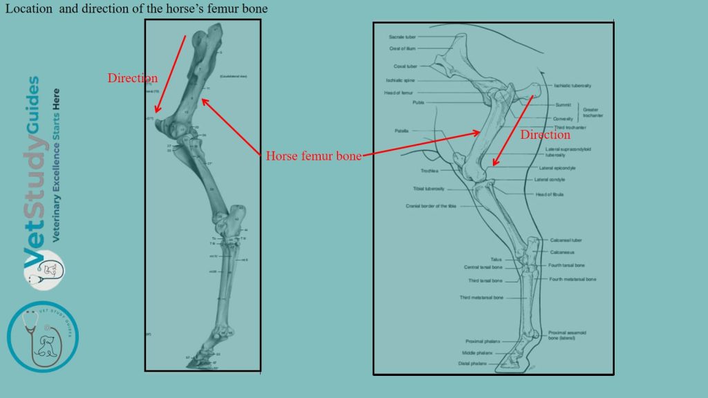
Horse femur bone
Type of bone: The horse’s femur or thigh bone, or os femoris, is the largest and most massive of the long bones of the skeleton.
Direction: It extends obliquely downward and forward, articulating with the acetabulum above and the tibia and patella below.
Description of horse femur bone anatomy
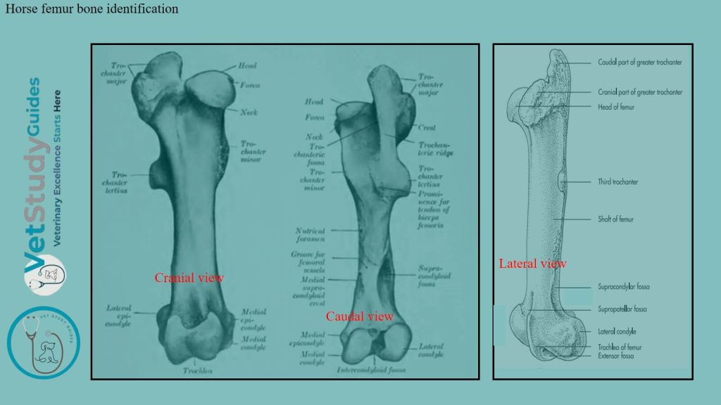
The horse femur presents for examination a shaft and two extremities.
Shaft/body/corpus femoris: it is cylindrical and presents anterior, posterior, lateral, and medial surfaces. It also presents the lateral and medial borders with different osteological features.
Two extremities: they are the proximal and distal extremities.
Shaft/body of the horse femur bone
The horse femur’s shaft/corpus femoris is, in general, cylindrical. But it is flattened behind, and larger above than below.
Anterior, medial, and lateral surfaces
Here, the anterior, medial, and lateral surfaces are continuous. They are strongly convex from side to side.
You will often find a central vertical rough line on the proximal part. However, most of these surfaces are smooth. They are covered by the quadriceps femoris muscle.
Posterior surface
The posterior surface of the femur’s shaft is wide, flat, and smooth in its proximal fourth. Distal to this part, there is a rough elevation laterally. It is for the attachment of the femoral tendon of the biceps femoris muscle.
However, there is a rough line medially to which the quadratus femoris is attached. The middle third is narrower and is rough for the attachment of the adductor muscle.
Just distal to this area, an oblique groove crosses the posterior surface. It indicates the position of the femoral vessels.
Medial border of the shaft
The medial border presents on its proximal part the trochanter minor. It is a thick, rough ridge to which the iho-psoas muscle is attached.
From this trochanter minor, a rough line curves up to the front of the neck. It indicates the posterior limit of the attachment of the vastus medialis muscle.
A narrow, rough area about the middle of the medial border gives attachment to the pectineus muscle. However, the nutrient foramen is usually found just in front of this border.
Supracondyloid crest: The medial supracondyloid crest is situated below the groove for the femoral vessels. It gives origin to the medial head of the gastrocnemius muscle of the hindlimb.

Lateral border of the femur’s shaft
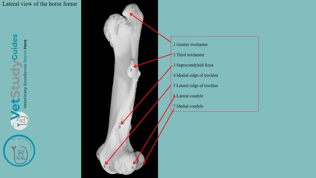
The lateral border of the shaft of the horse’s femur is prominent in its upper part. It bears at the junction of its proximal and middle thirds the trochanter tertius (third trochanter).
Here, this process is curved forward and has a thick edge. Within this edge, the tendon of the superficial gluteus muscle is attached.
Supracondyloid fossa: At the distal part is the supracondyloid fossa, in which the superficial digital flexor arises. It is bounded laterally by a thick, rough margin, the lateral supracondyloid crest. Within this crest, the lateral head of the gastrocnemius muscle is attached.
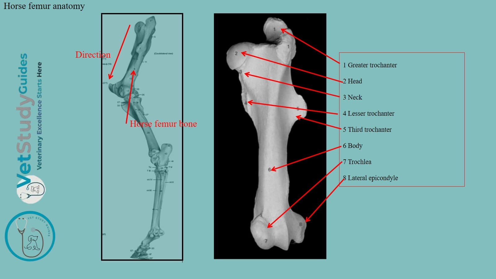
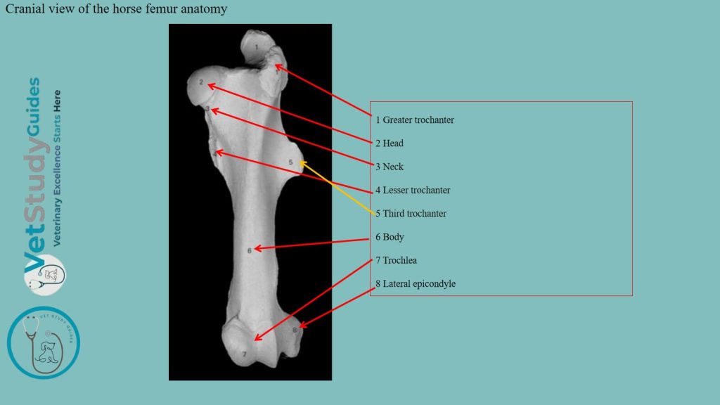
Proximal extremity of the horse femur
The proximal extremity/extremitas proximalis is large and consists of the head, neck, and trochanter major.
Head: The head/caput femoris is placed at the medial side. It is directed inward, upward, and somewhat forward.
Again, the head is approximately hemispherical and articulates with the acetabulum. It is cut intermediately by a deep notch, the fovea capitis.
Within the fovea capitis, the accessory and round ligaments of the femoral head are attached. A distinct margin surrounds the articular surface.
Neck: The neck/collum femoris is most distinct in front and medially. The trochanter major is situated laterally and presents three features.
The anterior part or convexity is situated opposite the head. It rises a little above the level of the latter. However, it gives attachment to the deep gluteus muscle.
In the fresh state, its lateral surface is coated with cartilage. Over this surface, a tendon of the middle gluteus passes and is inserted into the crest.
The posterior part or summit is separated from the anterior part by a notch. It is situated behind the plane of the head and rises to a much greater height.
However, it furnishes an insertion to part of the middle gluteus muscle. Its posterior border is continued downward as the trochanteric ridge.
Here, the trochanteric ridge forms the lateral wall of the trochanteric fossa. A number of foramina are found in the concave area medial to the convexity.
Distal extremity of the horse femur bone
The distal extremity/extremitas distalis of the horse femur is large in both directions. It comprises the trochlea in front and two condyles behind.
Trochlea: The trochlea consists of two ridges separated by a groove. They form an extensive surface/facies patellaris for articulation with the patella.
It is very unsymmetrical here; the medial ridge is much wider. However, the medial ridge is more prominent and extends higher than the lateral one.
Intercondyloid fossa: The condyles, medial and lateral, are separated by the deep intercondyloid fossa/fossa intercondyloidea. They articulate with the condyles of the tibia and the menisci of the stifle joint.
A ridge connects each condyle with the lower part of the corresponding ridge of the trochlea. The intercondyloid fossa lodges the spine of the tibia and the cruciate ligaments of the stifle joint, which are attached here.
Medial epicondyle: The medial epicondyle is a rounded prominence on the medial surface of the distal extremity. Within this structure, the collateral ligament and the adductor muscle are attached.
Lateral epicondyle: The corresponding lateral epicondyle is less distinct. It presents a mark where the lateral ligament is attached.
Below and behind the lateral ligament, there is a depression in which the popliteus muscle arises. Between the lateral condyle and trochlea is the extensor fossa.
Within this extensor fossa, the common tendon of origin of the extensor digitalis longus and peroneus tertius is attached.

FAQs on horse femur bone anatomy
The condyles are obliquely placed with their long axes directed downward, forward, and inward. The articular surface of the lateral condyle is more strongly convex from side to side than that of the medial one. Again, the ridge which connects it with the trochlea is much narrower.
The shaft and the distal end each ossify from one center. But the proximal end has two centers, one of which is for the head and the other for the trochanter major.
The edge of the trochanter tertius also has a separate center. The proximal end fuses with the shaft at three to three and a half years, the distal at about three and a half years.
Conclusion
So, the horse bone anatomy includes the description of the cylindrical shaft and two proximal extremities. Here, the shaft of a horse femur is unique as it contains a third trochanter.
The main features of the proximal extremity of the horse femur are the head, neck, and greater trochanter. However, the condyles and trochlea are important osteological features of its distal extremity.
References
- Sisson, S., Anatomy of the domestic animals. W B Saunders Company, USA.
- Dyce and Wensing, Textbook of Veterinary Anatomy, 4th edition, Saunders, USA.
- Anna Dee Fails and Christianne Magee, Anatomy and physiology of the Farm Animals, 111 River Street, Hoboken, NJ 07030, USA.
- He, H., Banks, S. A., & Biedrzycki, A. H. (2023). Anatomical variations of the equine femur. PloS one, 18(6), e0287381.
- Hilary M. Clayton, Peter F. Flood, Diana S. Rosenstein, and David Mandeville, Clinical Anatomy of the Horse, First edition 2005, ISBN 07234 3302 X.
- Pasquini and Spurgeon, Anatomy of domestic animals, systemic and regional approaches.
- Lang, J.J., Li, X., Micheler, C.M. et al. Numerical evaluation of internal femur osteosynthesis in the proximal equine hindlimb. BMC Vet Res 20, 188 (2024).
- Victoria Aspinall B, and Melanie Cappello, Introduction to Veterinary Anatomy and Physiology Textbook, ISBN 978-0-7020-5735-9, Elsevier.