
The horse thoracic vertebrae form the bony roof of the thoracic cavity. They are considered part of the axial skeleton and contribute to the formation of the vertebral canal.
Here, I will describe the osteological features of the horse’s thoracic vertebrae with diagrams.
What are the thoracic vertebrae in a horse?
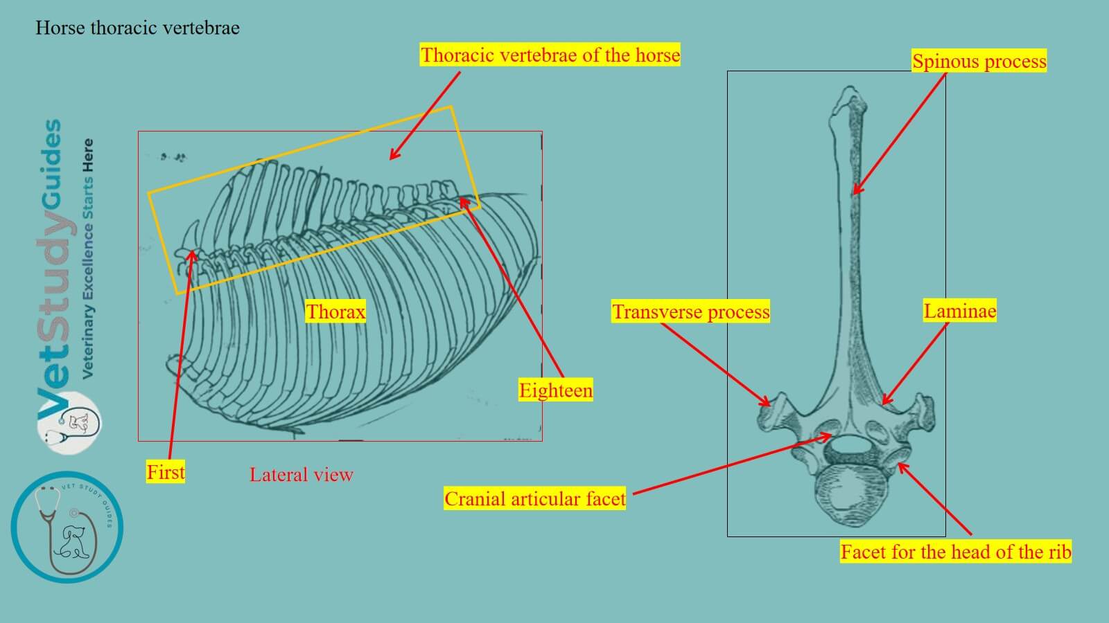
Here, the figure shows the thoracic vertebrae from the horse’s skeleton. These are the unpaired, irregular bones of the axial skeleton that form the bony roof of the thoracic region.

These thoracic vertebrae attach to the proximal extremity of the horse’s ribs. They have almost similar features to the typical vertebra of the horse. However, they are characterized by the presence of the long spinous process and cylindrical body.
Location of the thoracic vertebrae
Thus, the thoracic vertebrae are located in the dorsal part of the thorax or thoracic cavity. Here, the figure shows the location of the horse’s thoracic vertebrae from its skeleton.

Before describing the features of the horse’s thoracic vertebrae, you might have knowledge of a typical vertebra. However, the typical vertebra of a horse possesses one body, right and left lateral arches, and three processes.
Here, the processes are paired articular (cranial and caudal), paired transverse (right and left), and a single spinous process.
How many thoracic vertebrae are in a horse?
Number: The thoracic vertebrae or vertebrae thoracales are usually eighteen in number in the horse’s skeleton. However, there are sometimes nineteen, rarely seventeen, thoracic vertebrae.
Here, the figure shows eighteen thoracic vertebrae from the horse’s skeleton. However, the number of the horse’s thoracic vertebrae corresponds to the number of the ribs.

Horse thoracic vertebrae anatomy
In regional characters, the horse’s thoracic vertebrae articulate with the ribs. However, the spinous processes have variable length and form.
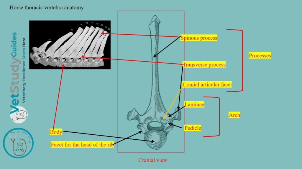
Those in the middle of the series are the most typical and present the following features –
- Body: They are cylindrical and possess paired cranial and caudal facets for the ribs.
- Arches: Pedicles are short, whereas the laminae are moderate in length. Like typical vertebrae, they also possess notches on the cranial and caudal aspects.
- Processes: Articular processes are not developed, whereas the spinous processes are longer. However, the transverse processes are short and bear facets for the ribs.
But I will describe the body, arches, and three processes in detail, with diagrams.
Body of the equine thoracic vertebrae
Shape and appearance: The bodies of the horse’s thoracic vertebrae are short and constricted in the middle.
Extremities: The extremities are expanded and have articular surfaces that are not strongly curved. Here, the anterior surface is convex, whereas the posterior surface is concave.
Facets: On the upper part of each side, there are anterior and posterior costal facets. They are also known as fovea costalis cranialis and caudalis.
Two facets of the adjacent vertebrae and the intervening fibro-cartilages form sockets for the heads of the ribs.

Arches of the horse thoracic vertebra
Like the typical vertebra, these thoracic vertebrae also have two lateral arches. However, these arches consist of the ventral pedicle and dorsal laminae.
Here, the arches of the horse’s thoracic vertebrae are small. Their posterior notches are relatively large and are often converted into foramina.

Thus, the anterior notch of the pedicles of the horse’s thoracic vertebrae is smaller. However, you will find the additional intravertebral foramina in the pedicles of the horse’s thoracic vertebrae.
Processes of the horse’s thoracic vertebrae
Like other vertebrae of the horse’s vertebral column, the thoracic vertebrae also possess three types of processes. They are the cranial and caudal paired articular surfaces, the paired lateral transverse surfaces, and the single dorsal spinous process.
Here, I will discuss these processes from the horse’s thoracic vertebrae with the diagrams.
Articular processes of the thoracic vertebrae
Small articular processes: The figure highlights the cranial and caudal articular processes from the horse’s thoracic vertebrae. Here, the articular processes of these thoracic vertebrae are small.
Anterior articular process: The anterior paired articular processes are, in fact, represented only by two oval facets. They are located on the anterior part of the arch, which face almost directly upward.
Posterior articular process: The posterior processes spring from the base of the spinous process. However, their facets face almost directly downward.

Transverse process of the thoracic vertebrae
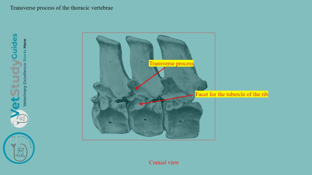
Appearances: The transverse processes of the equine thoracic vertebrae are short and thick. However, they are tuberous at the free end (laterally).
Facets: Each transverse process has a facet, called the fovea transversaria. They are for articulation with the tubercle of the rib, which has the same serial number.
Spinous process of the horse’s thoracic vertebrae
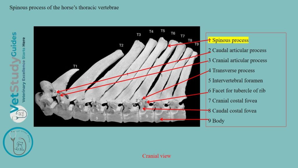
Unique features: Here, the spinous processes of the horse’s thoracic vertebrae show some unique features. The spinous process is large, narrow, and slopes upward and backward.
Borders: However, the anterior border of these spinous processes of the thoracic vertebrae is thin. Again, the posterior border is wider and furrowed.
Sometimes you will find exceptions at the summit of the spinous process. Here, the summit of the spinous process of the horse’s thoracic vertebrae is expanded and rough.
Features of the horse’s first thoracic vertebra
The first thoracic vertebra has the following specific characteristics –
Body of the first thoracic vertebra
The body of the first thoracic vertebra of a horse is wide and flattened dorso-ventrally. However, in front, it has a head like that of a cervical vertebra. The cavity is somewhat deeper than that of any other thoracic vertebra.
Costal facets: Two large costal facets are found on either side, and a well-marked spine ventrally.
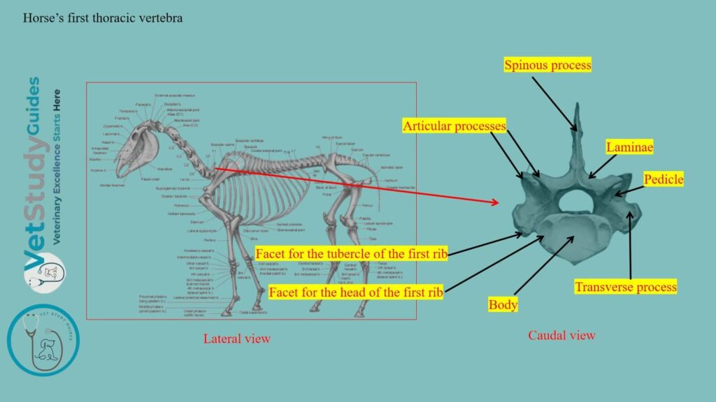
Arches and processes of the first thoracic vertebra
Arch: The arch of the horse’s first thoracic vertebra is large and strong, and has large notches.
Articular process: The articular processes are much larger than those of other thoracic vertebrae. They resemble those of the seventh cervical.
Transverse process: The transverse processes are short and thick in the first thoracic vertebra. However, each has on its ventral aspect a large concave facet for articulation with the tubercle of the first rib.
Spinous process: The spinous process is curved backward and tapers to a point. Its length is usually about three or four inches (8 to 10 centimeters).
Notes: However, this vertebra may be mistaken at first glance for the last cervical. Again, it is promptly identified by the three costal facets on each side and the length of the spine.
The last thoracic vertebra in the horse
The last thoracic vertebra in the horse is distinguished by the following features –
Absence of the posterior pair of costal facets, and
The confluence of the anterior pair with those on the transverse processes.
Serial position of the thoracic vertebrae in the horse’s skeleton
The serial position of other thoracic vertebrae may be determined at least approximately by the following data –
Features of the body
Length: The bodies gradually diminish in length and width to the mid of the vertebrae and then increase slightly.
Costal facets: Their costal facets become smaller and less concave from first to last. The ventral crest is distinct on three or four vertebrae at either end of the region.
Changes in the transverse processes
Size: The transverse processes diminish in size and are placed lower down as they are traced backward.
Costal facets: The costal facets of the transverse processes become smaller and lower. However, on the last (and sometimes on its predecessor also), it fuses with the costal facet of the body.
Mammillary process: The upper non-articular part of the process gradually becomes more sharply defined. However, in the last four or five forms, a distinct mammillary process.

Serial changes in spinous processes
Length: The spinous processes of the horse’s thoracic vertebrae increase in length to the third and fourth. However, then gradually diminish to the fifteenth, beyond which they have about the same length.
Inclination of the spinous process: The backward inclination is most pronounced in the second. However, in the sixteenth, it is vertical, and the last two are slightly forward . However, in the sixteenth, it is vertical, and the last two are slightly forward.
Longest spine: The longest spines (those of the withers) are the thickest. They have expanded summits that remain more or less cartilaginous. However, the others are more plate-like and are surmounted by a thick lip.
Second spine: The second spine is more than twice as large as the first. The summits of the fourth and fifth usually form the highest point of the withers.
FAQ’s on the horse’s thoracic vertebrae
Well, the horse’s thoracic vertebrae develop from the six or seven centers of ossification. However, three centers are for the body, two for the arch, and one for the spinous process. Some of the latter have an additional center for the summit.
Well, the body and spinous processes show exceptional features in the horse’s thoracic vertebrae compared to those of the ox. In the horse, the cranial border of the spinous process is thin, whereas the caudal border is divided into two areas.
Again, the few caudal thoracic vertebrae of the horse shows mammaliary processes on their spinous processes.
Conclusion
So, the horse thoracic vertebrae are the irregular bones of the axial skeleton. They also have the typical features of a vertebra, except for the spinous processes, transverse processes, and body.
Here, the horse’s thoracic vertebrae possess cranial and caudal facets for the ribs. However, the transverse process also possesses the facet for the articulation with the tubercle of the ribs. Again, the spinous processes are longer in the horse’s thoracic vertebrae compared to those of others.
References
- Sisson, S., Anatomy of the domestic animals. W B Saunders Company, USA.
- Dyce and Wensing, Textbook of Veterinary Anatomy, 4th edition, Saunders, USA.
- Spoormakers, T.J.P., et al. (2021). Comparative study of the anatomical configuration of the equine vertebra. J Anat, 239: 829-838.
- Beth Vanhorn and Robert W. Clark, Veterinary Assisting: Fundamentals & Applications, ISBN-13: 978-1-4354-5387-6, Maxwell Drive, Clifton Park, NY 12065-2919 USA.
- Anna Dee Fails and Christianne Magee, Anatomy and physiology of the Farm Animals, 111 River Street, Hoboken, NJ 07030, USA.
- Maciej Janeczek,Anatomical and Biomechanical Aspects of the Horse Spine, https://doi.org/10.1002/OA.2248
- Hilary M. Clayton, Peter F. Flood, Diana S. Rosenstein, and David Mandeville, Clinical Anatomy of the Horse, First edition 2005, ISBN 07234 3302 X.
- Haussler, Kevin K. Anatomy of the Thoracolumbar Vertebral Region, Veterinary Clinics: Equine Practice 15(1)
- Pasquini and Spurgeon, Anatomy of domestic animals, systemic and regional approaches.
- Victoria Aspinall B, and Melanie Cappello, Introduction to Veterinary Anatomy and Physiology Textbook, ISBN 978-0-7020-5735-9, Elsevier.
Reviewed and Edited by