Horse radius and ulna are the bones of the forelimb. Here, the horse’s radius possesses the typical features of a long bone, whereas the ulna is a reduced long bone in the horse.
In this guide, you will learn the osteological features of the horse radius and ulna bones separately.
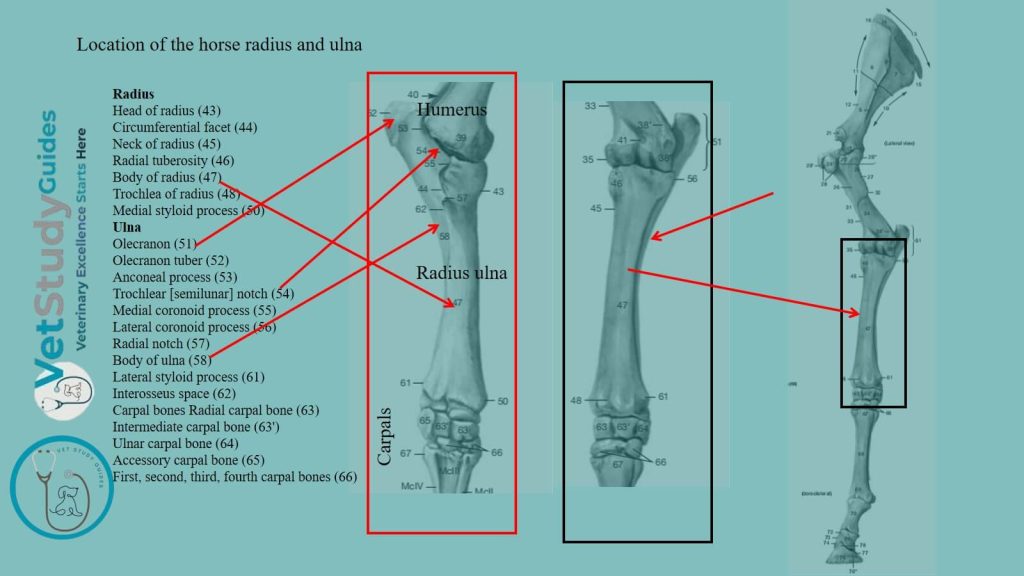
Horse radius bone
Location: The radius is much larger than the ulna of the forearm in the horse. It extends in a vertical direction from the elbow, where it articulates with the humerus, to the carpus.
However, the horse radius is gently curved, the convexity being dorsal. It consists of a body/shaft and proximal and distal extremities.
Shaft of the horse radius
The shaft/corpus radii is curved in its length. It is somewhat flattened from before backward and widened at its ends.
It presents for description two surfaces and two borders.

Surfaces of the shaft of the horse’s radius
Dorsal surface: The dorsal surface is smooth, slightly convex in its length, and rounded from side to side.
Volar/caudal surface: The volar surface is correspondingly concave in its length. It is flattened in the transverse direction.
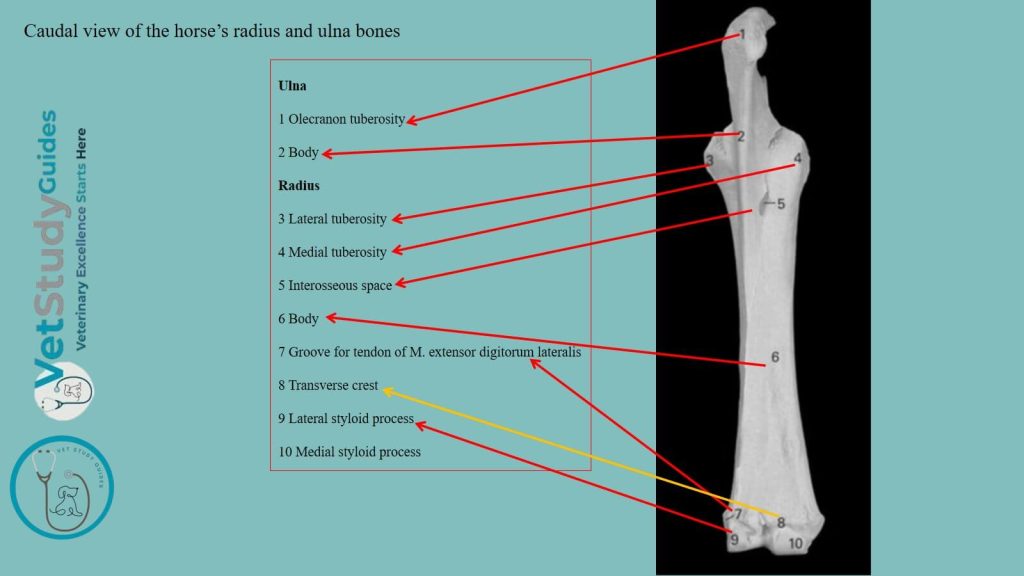
At its proximal part, there is a smooth, shallow groove. It concurs with the ulna in the formation of the interosseous space of the forearm. It also contains the nutrient foramen in the lower part of this groove.
Below this, there is in the young subject a narrow, rough, triangular area. Within this area ulna is attached by an interosseous ligament. In the older horse, the two bones (the horse’s radius and ulna) are fused here. A variable rough elevation distal to the middle and close to the medial border gives attachment to the radial check ligament.
Borders of the radius bone
Medial border: The medial border is slightly concave in its length and is largely subcutaneous. At its proximal end, there is a smooth area on which the tendon of insertion of the brachialis muscle lies.
A small rough area just below gives attachment to that muscle and the long medial ligament of the elbow joint.
Lateral border: The lateral border is more strongly curved, but presents no special features.
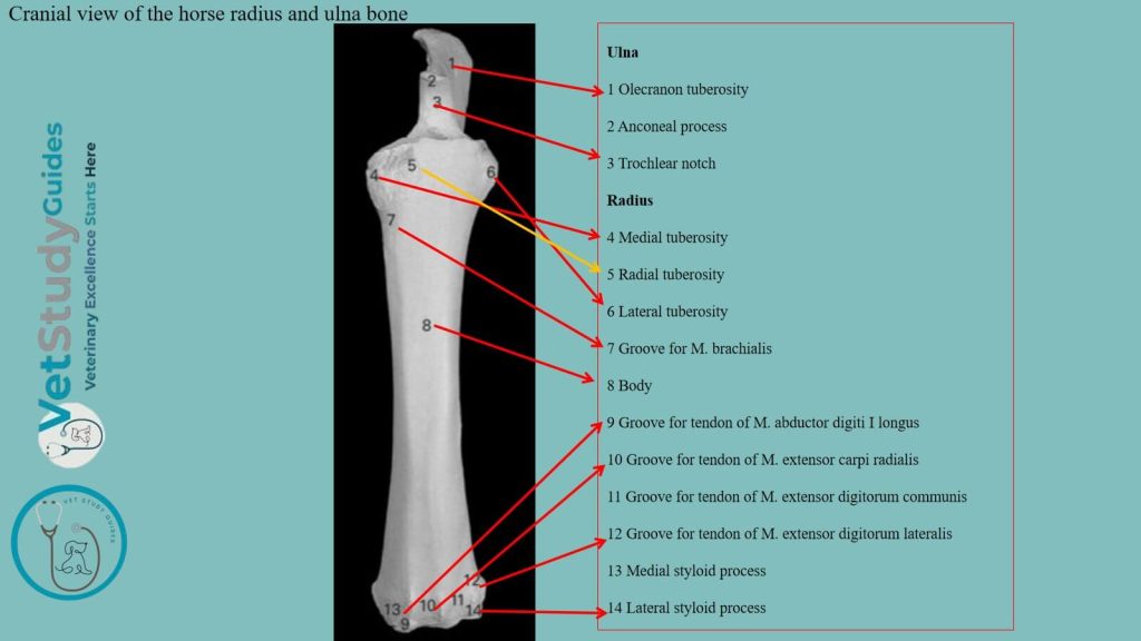
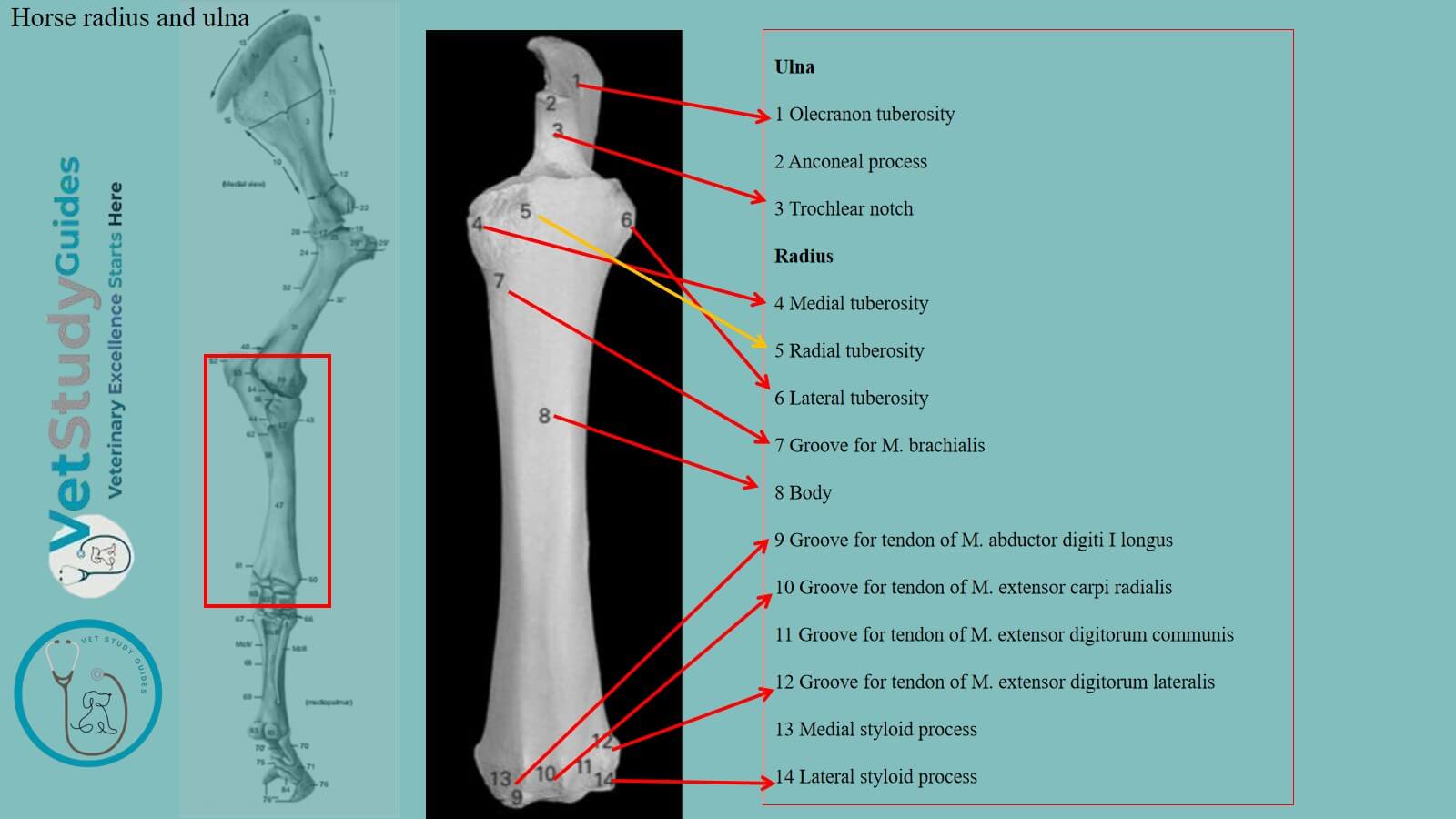
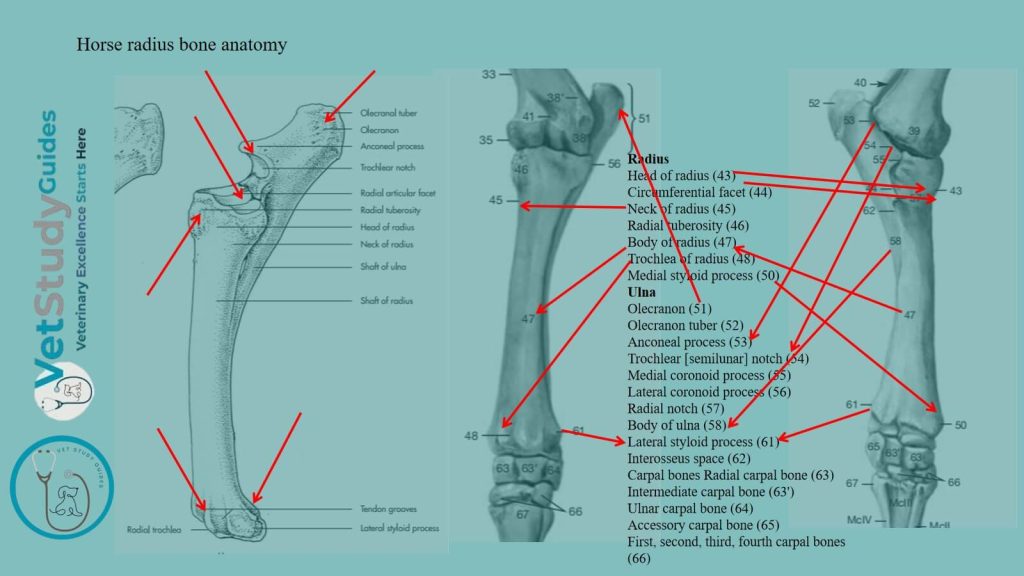
Proximal extremity of the horse radius bone
The proximal extremity or head is flattened from before backward and wide transversely. It presents the humeral articular surface. It corresponds to that of the distal end of the humerus;
This structure is crossed by a sagittal ridge, which has a synovial fossa on its posterior part. Again, it contains a prominent lip, the coronoid process at the ends in front.
Just below the posterior border, and separated by a depression, there are two concave facets. They are for articulation with the ulna.
However, the horses’ radius and ulna bones are united by an interosseous ligament.
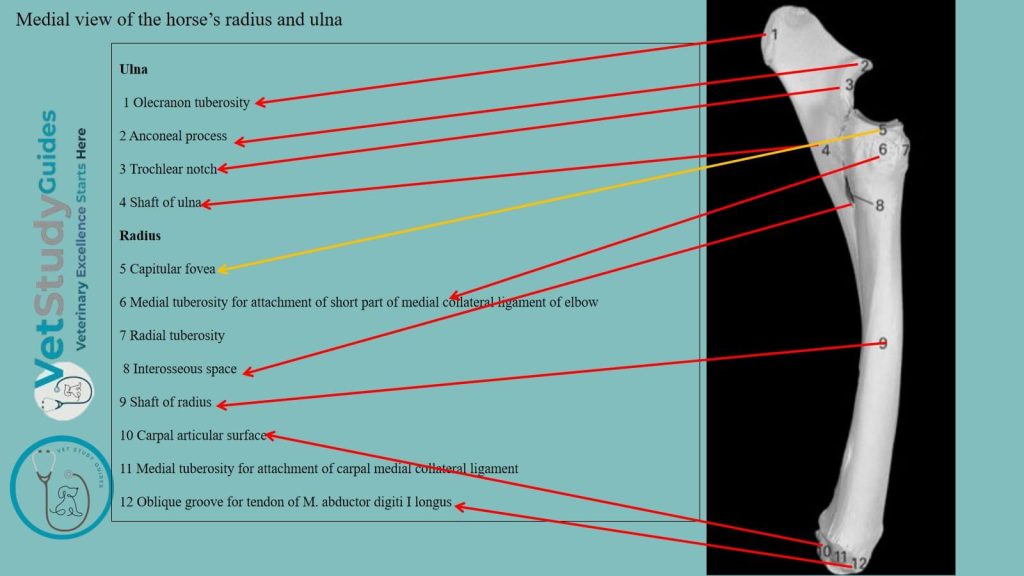
At the medial side of the dorsal surface is the radial/bicipital tuberosity. Within this tuberosity, the biceps tendon is inserted.
Medial tuberosity: The medial tuberosity is continuous with the preceding eminence. It furnishes attachment to the small part of the medial ligament.
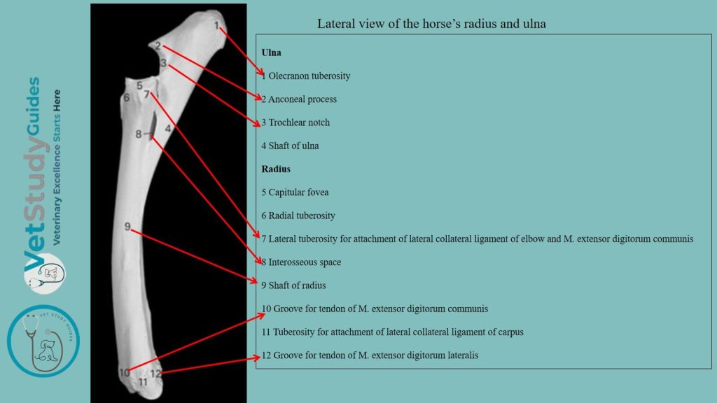
Lateral tuberosity: The lateral tuberosity is more salient. It gives attachment to the lateral ligament and to the common and external extensor muscles of the digit.
Distal extremity of the horse radius bone
The distal extremity is also compressed from before backward. It presents the carpal articular surface, which consists of three parts.
Medial facet: The medial facet is the largest, is quadrilateral, concavo-convex from before backward. It articulates with the radial carpal bone.
Intermediate facet: The intermediate one is somewhat similar in form but smaller. It articulates with the intermediate carpal bone.
Lateral facet: The lateral facet is smaller, convex, and articulates below with the ulnar carpal and behind with the accessory carpal.
Surfaces from the distal extremity of the horse radius
The dorsal surface presents three grooves, separated by ridges.
Middle groove: The middle one is vertical and gives passage to the tendon of the extensor carpi radialis.
Lateral groove: The lateral one is similar and provides the tendon of the common digital extensor.
Medial groove: The medial one is small and oblique and lodges the tendon of the extensor carpi obliquus.
The volar aspect is crossed by a rough ridge, below which are three depressions. On either side is a tuberosity to which the collateral ligament is attached.
A small vertical groove marks the lateral one for the passage of the lateral extensor tendon.
Now, we will discuss the osteological features of the horse ulna bone.
Horse ulna bone anatomy
Location and type of bone: The ulna of the horse is a reduced long bone situated behind the radius. The radius, it is partially fused in the older horse.
The horse radius bone also possesses the shaft and two extremities for description.
The shaft of the horse ulna
The shaft is three-sided and tapers to a point distally.
Dorsal and volar surfaces: The dorsal surface is applied to the volar surface of the radius. Just below the interosseous space, the horse’s radius and ulna bones are fused in the older horses.
The surface which enters into the formation of the space is smooth. It usually presents a small nutrient foramen, directed upward.
Above the space, it is rough and is attached to the radius by an interosseous ligament, which is usually permanent.
Medial and lateral surfaces: The medial surface is smooth and slightly concave. Again, the lateral surface is flattened.
Medial and lateral borders: The medial and lateral borders are thin and sharp, except at the interosseous space.
Volar border: The volar border is slightly concave in its length and is rounded. Distal end: The distal end is pointed and is usually a little below the middle of the radius. A fibrous cord commonly continues to the distal tuberosity of the radius. But this band may be replaced in part or entirely by bone.
Extremities of the horse ulna
Proximal extremity: The proximal extremity is the major part of the bone. It projects upward and somewhat backward behind the distal end of the humerus. It forms a lever arm for the extensor muscles of the elbow.
Surfaces: The medial surface is concave and smooth. The lateral surface is convex and is roughened above.
Dorsal borders: The dorsal border bears on its middle a pointed projection. It is known as the processus anconaeus or beak. Here, the overhangs the semilunar notch of the horse radius.
The latter is triangular in outline, concave from above downward, convex transversely. It articulates with the humerus.
In its lower part, there is an extensive synovial fossa. Just below the notch are two convex facets which articulate with those on the volar aspect of the proximal end of the radius.
Volar border: The volar border is nearly straight and is thick and rounded. The free end or summit is a rough tuberosity, the olecranon, which gives attachment to the triceps brachii and other muscles.
Distal extremity: The distal extremity has fused with the radius.
FAQ’s on horse radius and ulna bones
The radius ossifies from four centers, viz., one each for the shaft, the two extremities, and the lateral part of the distal end; the last is morphologically the distal end of the ulna, which has fused with the radius, and a distinct groove on the carpal articular surface often indicates the line of fusion. The proximal extremity unites with the shaft at about one and a half years, the distal end at about three and a half years, usually.
The horse ulna ossifies from three centers. Here, one is for the main part of the bone, one for the olecranon, and one for the distal end.
The cartilaginous embryonic ulna extends the entire length of the forearm. The distal part of the shaft is usually reduced to a small fibrous band or may disappear entirely.
The distal extremity fuses before birth with the radius. The olecranon unites with the rest of the bone at about three and a half years. A medullary cavity appears to occur constantly in the adult.
Conclusion
So, the horse radius and ulna bones possess unique osteological features on their shaft and extremities. Here, the horse radius is the larger cranial, and the ulna is the smaller reduce long bone that attaches to the caudolateral aspect of the radius.
References
- Sisson, S., Anatomy of the domestic animals. W B Saunders Company, USA.
- Rogers, C. W., Gee, E. K., & Dittmer, K. E. (2021). Growth and Bone Development in the Horse, Animals, 11(12), 3402.
- Dyce and Wensing, Textbook of Veterinary Anatomy, 4th edition, Saunders, USA.
- de Alcântara Leite dos Reis, D., Gouveia, B.L.R., Júnior, J.C.R. et al. Comparative evaluation of anatomical details of thoracic limb bones of a horse to those of models produced via scanning and 3D printing. 3D Print Med 5, 13 (2019).
- Anna Dee Fails and Christianne Magee, Anatomy and physiology of the Farm Animals, 111 River Street, Hoboken, NJ 07030, USA.
- Hilary M. Clayton, Peter F. Flood, Diana S. Rosenstein, and David Mandeville, Clinical Anatomy of the Horse, First edition 2005, ISBN 07234 3302 X.
- Pasquini and Spurgeon, Anatomy of domestic animals, systemic and regional approaches.
- Solounias, N., Danowitz, M., Stachtiaris, E., Khurana, A., Araim, M., Sayegh, M., & Natale, J. (2018). Evolutionary anatomy of the horse’s manus. Royal Society Open Science, 5(1), 171782.
- Victoria Aspinall B, and Melanie Cappello, Introduction to Veterinary Anatomy and Physiology Textbook, ISBN 978-0-7020-5735-9, Elsevier.