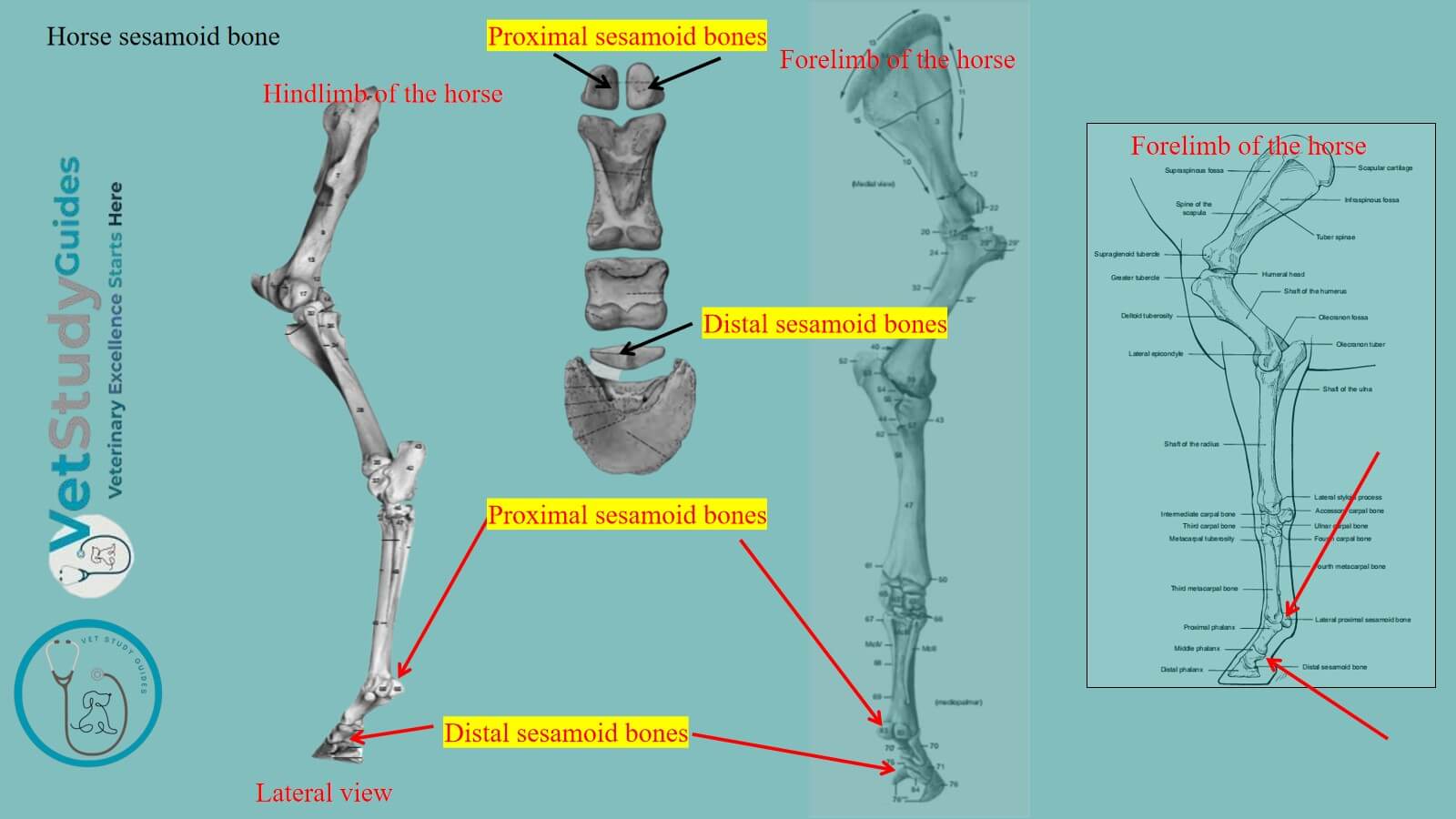
The horse sesamoid bone is a small, seed-like bone develop within the tendon and ossifies after birth. Here, I will describe the different types of sesamoid bones from the horse skeleton.
Sesamoids found in the phalanges of the horse’s forelimb and hind limb are a small type of bone. However, the horse patella is considered the largest sesamoid bone.
Types of sesamoid bones in a horse
There are two types of small sesamoid bones in the horse’s limb –
- Proximal sesamoids (2 in number in each digit), and
- Distal sesamoid (1 in number in each digit).
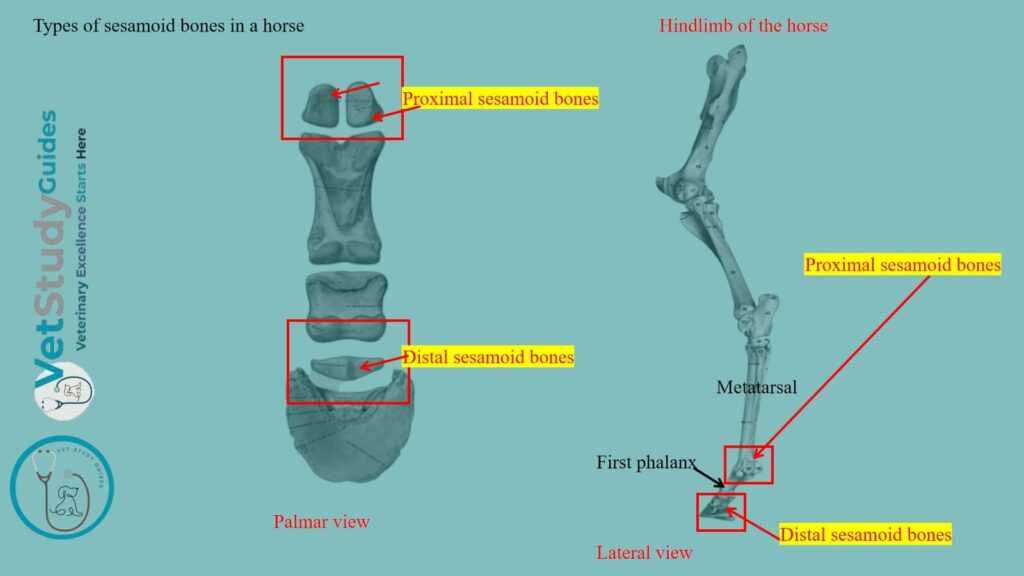
Table 1 shows the types and number of the horse’s sesamoids in their forelimb and hindlimb –
| Type of sesamoids | Number in each limb | Total numbers |
| Proximal sesamoids | 2 (two) | 8 (eight) |
| Distal sesamoids | 1 (one) | 4 (four) |
Where is the sesamoid bone in a horse?
The horse’s sesamoid bones/ossa sesamoidea proximalia are located just proximal to the fetlock joint on the palmar/planter aspect of the corresponding limbs.
They are shaped like a three-sided pyramid with their apex pointing proximally. However, they are firmly attached, and strong ligaments hold the first phalanx.
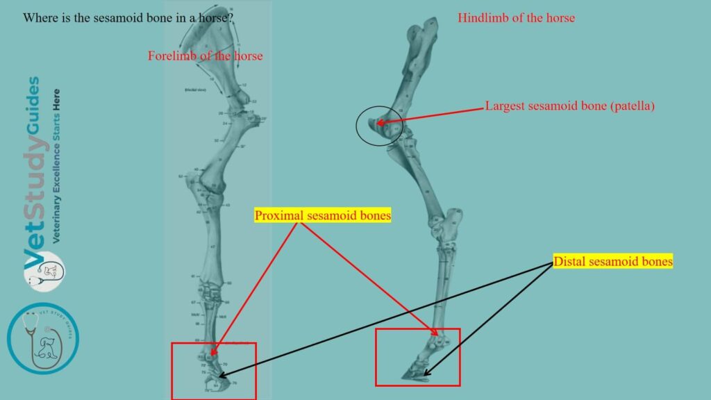
Table 2 shows the location of the horse’s proximal and distal sesamoid bones –
| Type of horse’s sesamoid | Location of the sesamoids |
| Horse’s proximal sesamoids | Palmar/planter aspects of the proximal and middle phalanges |
| Horse’s distal sesamoids | Palmar/planter aspects of the middle and distal phalanges |
Proximal sesamoids in the horse
Location and numbers: The two proximal sesamoids/ossa sesamoidea phalangis primse are situated behind the distal end of the large metacarpal bone. They are closely attached to the first phalanx by strong ligaments.
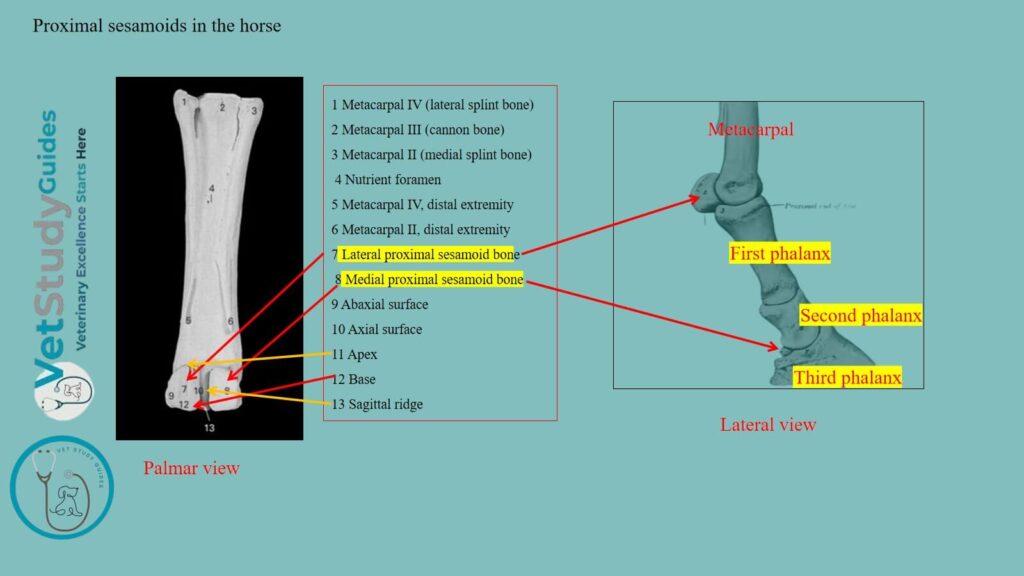
Shape: Each has the form of a three-sided pyramid.
Description: For description purpose, the sesamoid of a horse has 3 surfaces (articular, flexor, and abaxial), a base, and an apex.
Articular surface: The articular surface or facies articularis conforms to the corresponding part of the distal end of the large metacarpal bone.
Flexor surface: The flexor surface or facies flexoria is flattened and oblique. In the fresh state, it is covered by a layer of cartilage.
It also fills the interval between the opposed borders of the two bones. However, it forms a smooth groove for the deep flexor tendon.
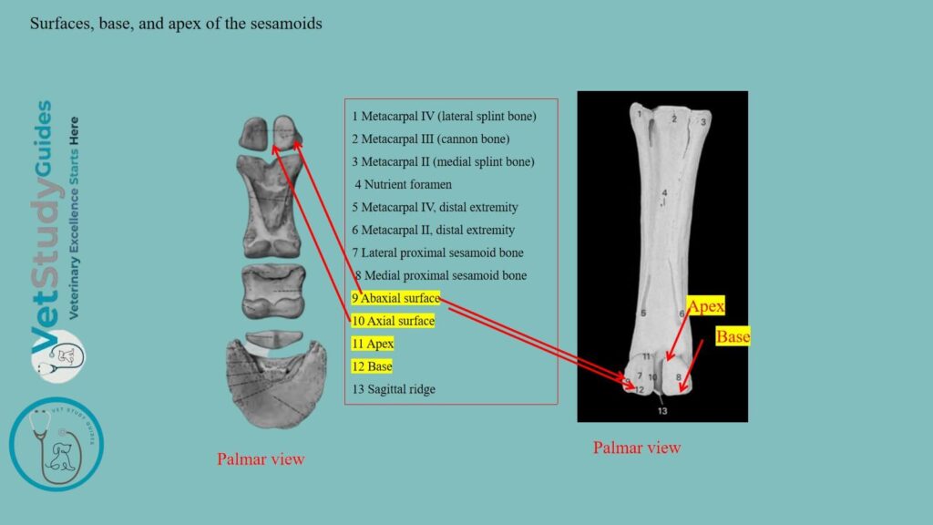
Abaxial surface: The abaxial surface is concave and gives attachment to part of the suspensory ligament. It is separated from the flexor surface by a rough everted border.
Base and apex: The base is distal and furnishes attachment to the distal sesamoidean ligaments. However, the apex is proximal and is rounded.
Distal sesamoid bones in the horse
Location and numbers: The distal sesamoid or navicular bone or os sesamoideum phalangis tertise is shuttle-shaped. It is situated behind the junction of the second and third phalanges.
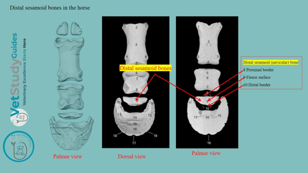
Description: Its long axis is transverse, and it possesses two surfaces, two borders, and two extremities.
Articular surface: The articular surface or facies articularis faces upward and forward. It consists of a central eminence, flanked by concave areas.
However, it articulates with the distal end of the second phalanx.
Flexor surface: The flexor or tendon surface or facies flexoria is directed downward and backward. It resembles the articular surface in form, but is more extensive and not so smooth.
In the fresh state, it is coated with cartilage, and the deep flexor tendon passes over it.
Proximal border: The proximal border or margo proximalis is wide and grooved in its middle. However, it is narrower and rounded on either side.
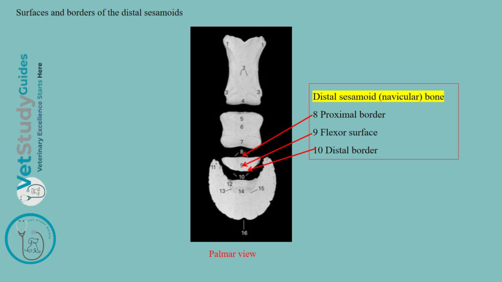
Distal border: The distal border or margo distalis bears in front a narrow facet for articulation with the third phalanx. Behind this is a groove, which contains a number of relatively large foramina.
Again, it is bounded behind by a prominent edge.
Extremities: There are two extremities which are blunt-pointed. Each of the sesamoids of the horse ossifies from a single center.
FAQs on the horse sesamoids
The upper and lower divisions of the angle of the sesamoids are sometimes termed the basilar and retrossal processes, respectively. These are usually called the lateral cartilages, but this designation could not be retained.
Conclusion
So, the horse sesamoid bones are the smallest bones in the fetlock joints of both forelimbs and hindlimbs. There are a total of eight proximal sesamoid bones in the 4 limbs of the horse. However, there are total four distal sesamoid bones in the limbs of the horse.
References
- Sisson, S., Anatomy of the domestic animals. W B Saunders Company, USA.
- Dyce and Wensing, Textbook of Veterinary Anatomy, 4th edition, Saunders, USA.
- Cornelissen BP et al., Nerve supply of the proximal sesamoid bone in the horse. Vet Q. 1994 May;16 Suppl 2:S66-9.
- Beth Vanhorn and Robert W. Clark, Veterinary Assisting: Fundamentals & Applications, ISBN-13: 978-1-4354-5387-6, Maxwell Drive, Clifton Park, NY 12065-2919 USA.
- Anna Dee Fails and Christianne Magee, Anatomy and physiology of the Farm Animals, 111 River Street, Hoboken, NJ 07030, USA.
- Pares-Casanova, Morphometry of equine distal sesamoid bone on clinically normal horses. J Dairy Vet Anim Res. 2015;2(1):19‒22.
- Elane, G. L. et al., (2022). Kinematics of the equine distal sesamoid (navicular) bone of the thoracic limb. American Journal of Veterinary Research, 83(7), Article ajvr.21.07.0090, ajvr.21.07.0090. Retrieved Jun 3, 2026, from
- Hilary M. Clayton, Peter F. Flood, Diana S. Rosenstein, and David Mandeville, Clinical Anatomy of the Horse, First edition 2005, ISBN 07234 3302 X.
- Pasquini and Spurgeon, Anatomy of domestic animals, systemic and regional approaches.
- Victoria Aspinall B, and Melanie Cappello, Introduction to Veterinary Anatomy and Physiology Textbook, ISBN 978-0-7020-5735-9, Elsevier.
Reviewed and Edited by