
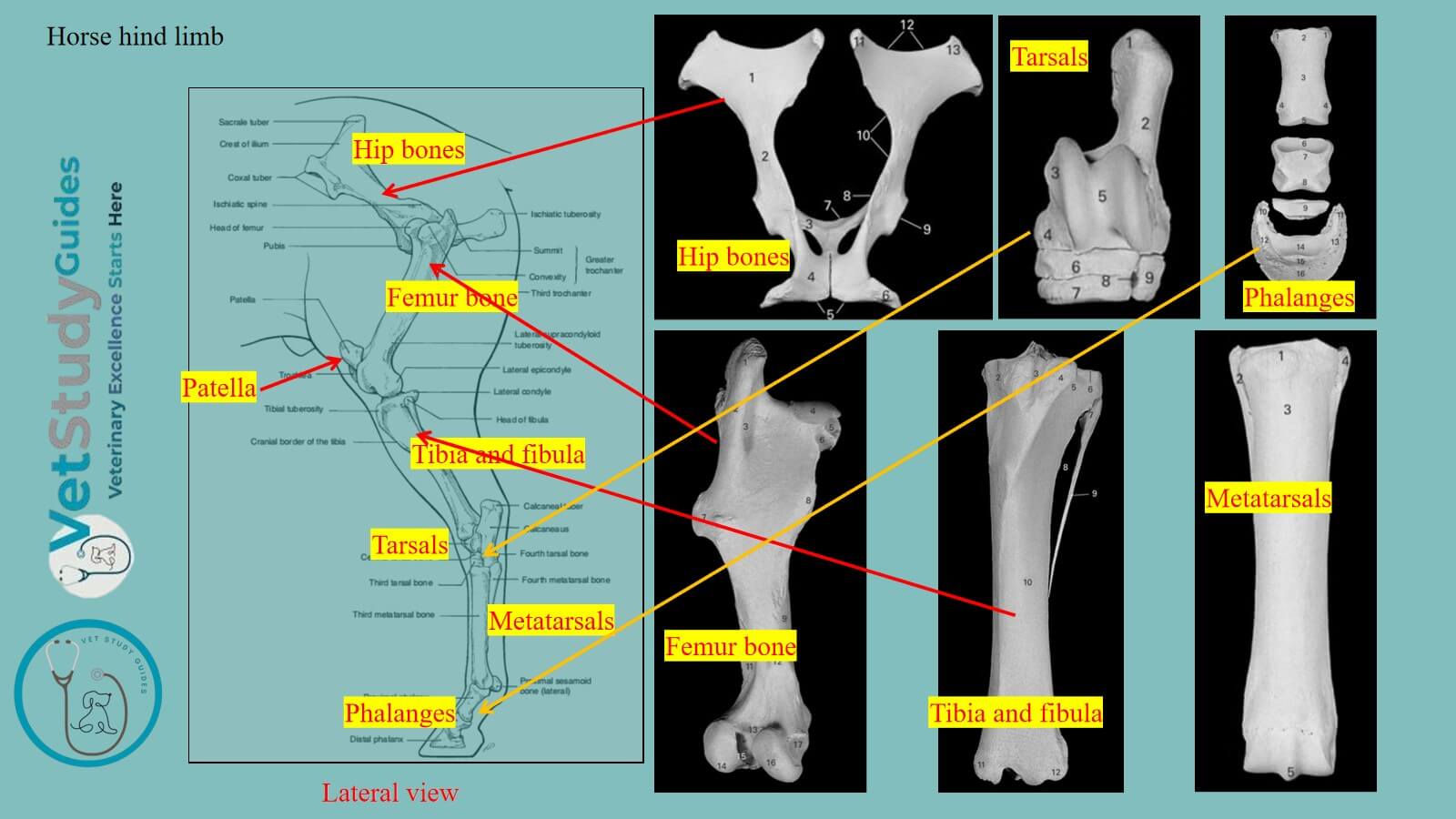
Horse hind limb forms part of the appendicular skeleton and consists of different bones. Here, I will describe the osteological features of the different bones from the horse’s hind limb with diagrams.
Like the segments of the horse thoracic limb, you will also find various segments in the hind/pelvic limb. Thus, I will also describe the segments of the hind limb along with their contents.
Horse hind limb skeleton
The horse’s hind/pelvic limb shows four different main segments. So, first let’s identify these four main segments from the hind limb –
- First segment: the pelvic girdle,
- Second segment: thigh,
- Third segment: leg, and
- Fourth segment: the pes
Here, the last segment, or pes, is subdivided into the tarsus, metatarsus, and digits. However, the figure identifies the various segments of the horse’s hind limb along with their bones.

Bones of the horse’s pelvic/hind limb
The figure shows that each segment of the hind limb contains single or multiple bones. Thus, you might first identify these bones and learn their osteological features thoroughly.

Here, the horse’s hind limb consists of the following bones –
- Hip bones or pelvic bones (right and left; each consists of ilium, ischium, and pubis): They form the bony pelvis/ pelvic girdle in the horse.
- Femur bone: It is a long, cylindrical, or round bone of the hind limb and forms the thigh segment.
- Tibia and fibula: They are also modified long bones that form the leg segment of the hind limb.
- Tarsal bones: They are the small bones that form the tarsus in the hind limb.
- Metatarsal bones: There are two types (large and small) and form the metatarsus region in the hind limb.
- Phalanges: There are three developed phalanges in each hind limb and form a single developed digit.
Here, the tarsals, metatarsals, and phalanges contribute to forming the pes segment of the hind limb. Now, I will describe these bones from the horse’s hind limb with a diagram.
Pelvic girdle of the horse
The pelvic girdle/cingulum extremitatis pelvinse of the horse consists of the os coxae or hip bone. They join their fellow of the opposite side ventrally at the symphysis pubis.
However, they also articulate very tightly with the sacrum dorsally. However, the part that joins with the sacrum is known as the tuber sacrale.

Bony pelvis: The two coxal bones, together with the sacrum and the first three or more coccygeal vertebrae, constitute the bony pelvis.
Bones of the os coxae: The os coxae consists originally of three flat bones, the ilium, ischium, and pubis. These bones meet at the acetabulum, a large cotyloid cavity with which the head of the femur articulates.
These three parts are fused before growth is complete, but are considered separately for the convenience of description.
- Ilium bone: The ilium/os ilium is situated in the lateral wall of the pelvis.
- Pubis bone: The pubis/os pubis is in the anterior part.
- Ischium: The ischium/os ischii in the posterior part of the ventral wall.
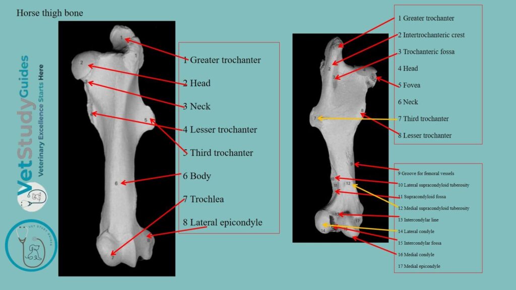
Horse thigh bone or femur
The thigh/femur of the horse, like the arm, contains a single long bone. It is known as the femur or thigh bone, or os femoris.
Articulation: This bone articulates with the acetabulum above and the tibia and patella below.
Horse leg bones/ tibia and fibula
The skeleton of the horse’s leg/crus comprises three bones: the tibia, fibula, and patella.
Tibia bone: The tibia is a large, prismatic long bone that supports the weight. It articulates distally with the tibial tarsal bone of the horse’s hind limb.
Fibula bone: The fibula is situated along the lateral border of the tibia. However, this bone is separated from the tibia by the interosseous space of the leg.
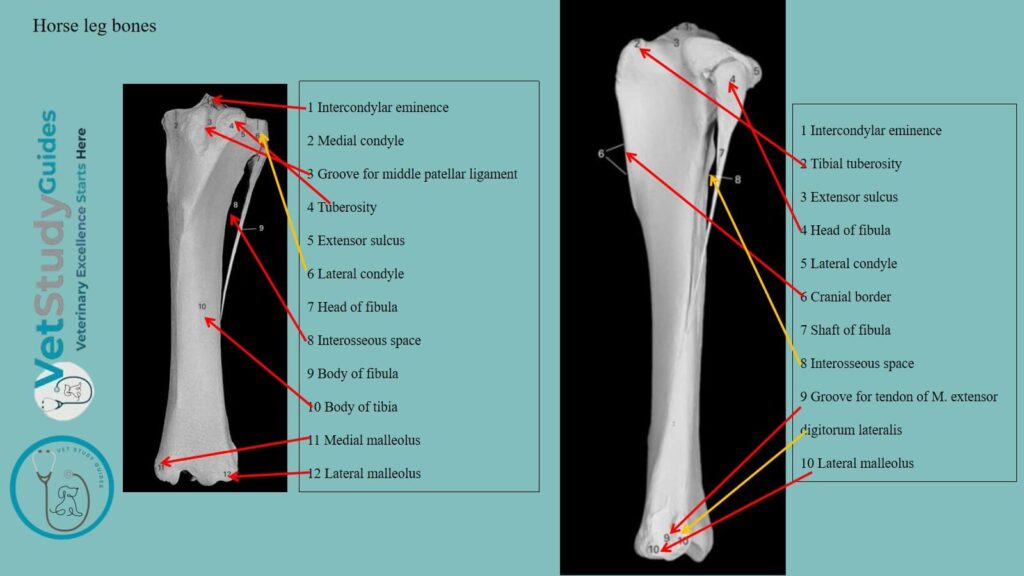
Exceptions: It (fibula) is much more slender than the tibia and does not articulate with the femur.
Pig and dog tibia and fibula bones: In the pig and dog, they have a complete shaft and two extremities. However, in the horse and ox, it is much reduced and otherwise modified.
Horse knee cap/ patella bone
The patella of the horse is a short bone that articulates with the trochlea of the distal end of the femur. It is also known as the knee-cap in horses.
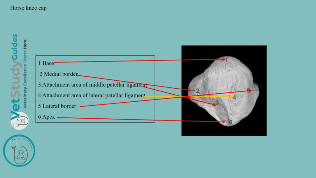
However, it is to be regarded as a large sesamoid bone intercalated in the tendon of the quadriceps femoris muscle.
Horse tarsus or hock
The tarsus or hock of the horse consists of a group of short bones, the ossa tarsi. There are 5 to 7 tarsal bones in each animal.
Proximal tarsals: In the horse, the proximal or crural row consists of two bones, the tibial and fibular tarsals. However, the former is situated at the tibial side and has a trochlea for articulation with the distal end of the tibia.
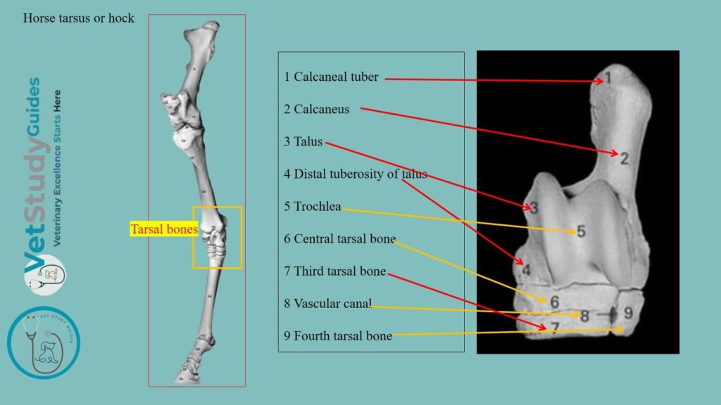
Again, the latter, situated at the fibular side, has a process, the tuber calcis. It projects upward and backward and constitutes a lever for the muscles that extend the hock joint.
Distal tarsals: The distal or metatarsal row consists of four bones when seven tarsal elements are present, as in the pig and dog. They are best designated numerically as first tarsal, second tarsal, etc.
Middle or central tarsal: The central tarsal is interposed between the rows. The Latin names and synonyms are given below (In Table 1) –
| Name of tarsal bone | Anatomical name | Symbol |
| Tibial tarsal | Os tarsi tibiale | Tt |
| Astragalus or Talus Fibular (Os tarsi fibulare, Tf.) | Astragalus or Talus Fibular (Os tarsi fibulare, Tf.) | Tf |
| Calcaneus or Os calcis Central (Os tarsi centrale, Tc.) | Calcaneus or Os calcis Central (Os tarsi centrale, Tc.) | Tc |
| Scaphoid or Navicular First Tarsal (Os tarsale primum, Tl) | Scaphoid or Navicular First Tarsal (Os tarsale primum, Tl) | T1 |
| First or internal cuneiform | Os tarsale secundum | T2 |
| Second or middle cuneiform | Os tarsale secundum | T2 |
| Third Tarsal or external cuneiform | Os tarsale tertium | T3 |
| Fourth Tarsal or Cuboid | Os tarsale quartum | T4 |
Horse metatarsals and phalanges
The metatarsal and digital bones of the horses resemble those of the corresponding regions of the thoracic limb. However, a few differential features are noted in the special descriptions.
There is one large (III) metatarsal and two small metatarsals (II and IV) present in the horse’s hind limb. Here, the small metatarsals articulate with the large ones on either side of the posterior aspect.
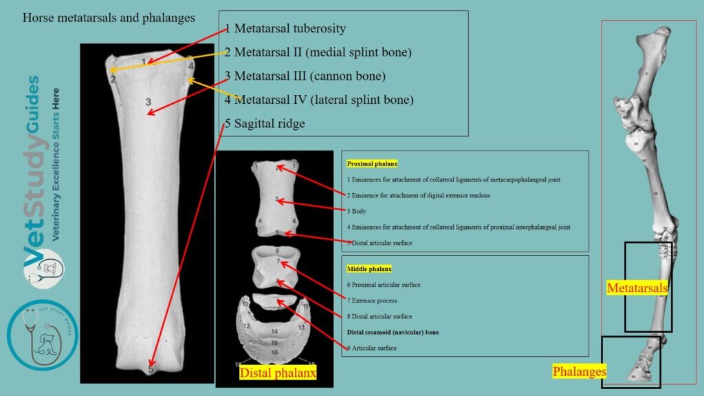
However, the large metatarsal of a horse is longer than the large metacarpal. Again, its proximal extremity is also wider than that of the large metacarpal bone.
However, the phalanges of the horse’s hind limb are almost similar to those of the forelimb or thoracic limb.
FAQ’s on the horse hind limb skeleton
Well, there are various distinguishing features of the hind limbs of the horse and the ox. Here, the ox coxae are wider in the horse than in the ox. However, the horse’s femur possesses a third trochanter, which is absent in the ox femur.
The fibula bone does not reach the distal end in the horse. However, the tarsus and metatarsals show great variation in their number and appearance in the horse compared to the ox.
Conclusion
So, the horse hind limb skeleton is formed by the bones of the hip, femur, tibia, fibula, tarsals, metatarsals, and phalanges. Here, the osteological features of these bones are unique from those of other species.
However, there are four different segments in the horse’s hind limb. Bones of the hind limb form these different segments and are termed the pelvic girdle, thigh, leg, and pes.
References
- Sisson, S., Anatomy of the domestic animals. W B Saunders Company, USA.
- Chamadia R et al., Decoding hip joint anatomy on radiographs and magnetic resonance imaging. Indian J Musculoskelet Radiol. 2025;7:3-12.
- Dyce and Wensing, Textbook of Veterinary Anatomy, 4th edition, Saunders, USA.
- Vanhorn, B., and Clark, R. W. Veterinary Assisting: Fundamentals & Applications. ISBN-13: 978-1-4354-5387-6. Maxwell Drive, Clifton Park, NY 12065-2919, USA.
- Fails, A. D., and Magee, C. Anatomy and Physiology of the Farm Animals. 111 River Street, Hoboken, NJ 07030, USA.
- Fürst, A., Meier, D., Michel, S., Schmidlin, A., Held, L., & Laib, A. (2008). Effect of age on microarchitecture in the radius and tibia of horses: an Xtreme computed tomographic study. BMC Veterinary Research, 4, 3.
- Pasquini and Spurgeon, Anatomy of domestic animals, systemic and regional approaches.
- The metacarpal/metatarsal region. In Equine MRI, R.C. Murray (Ed.) by Brokken, (2010). https://doi.org/10.1002/9781118786574.ch7
- Hilary M. Clayton, Peter F. Flood, Diana S. Rosenstein, and David Mandeville, Clinical Anatomy of the Horse, First edition 2005, ISBN 07234 3302 X.
- Aspinall and Cappello, M. Introduction to Veterinary Anatomy and Physiology Textbook. ISBN 978-0-7020-5735-9. Elsevier.
- Banks, Anatomical features of the equine femur and tibia bones. 2023; PLOs, 18(6), 0287381.
Reviewed and Edited by